. 2008 Jun 16;181(6):999-1012.
doi: 10.1083/jcb.200710187.

Regulation of RhoA-dependent ROCKII activation by Shp2

Affiliations

Regulation of RhoA-dependent ROCKII activation by Shp2

Hsiao-Hui Lee et al. J Cell Biol. .

Abstract

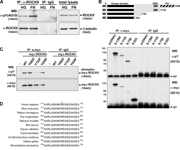
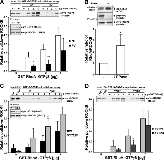
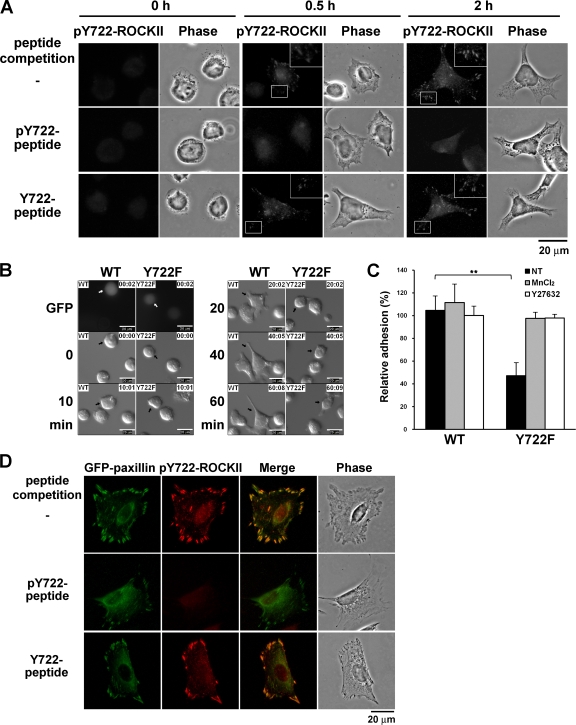
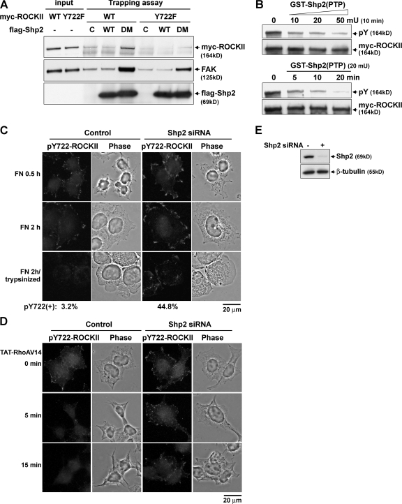
Contractile forces mediated by RhoA and Rho kinase (ROCK) are required for a variety of cellular processes, including cell adhesion. In this study, we show that RhoA-dependent ROCKII activation is negatively regulated by phosphorylation at a conserved tyrosine residue (Y722) in the coiled-coil domain of ROCKII. Tyrosine phosphorylation of ROCKII is increased with cell adhesion, and loss of Y722 phosphorylation delays adhesion and spreading on fibronectin, suggesting that this modification is critical for restricting ROCKII-mediated contractility during these processes. Further, we provide evidence that Shp2 mediates dephosphorylation of ROCKII and, therefore, regulates RhoA-induced cell rounding, indicating that Shp2 couples with RhoA signaling to control ROCKII activation during deadhesion. Thus, reversible tyrosine phosphorylation confers an additional layer of control to fine-tune RhoA-dependent activation of ROCKII.


Figures

Figure 1.
Figure 1.
Involvement of tyrosine dephosphorylation in ROCK-dependent cell rounding. (A–C) Adherent D2 cells plated in culture dishes in serum-free medium for 2 h were serum stimulated and treated with 300 μM orthovanadate (Na3VO4) or 20 μM Y27632, followed by stimulation with 20% heat-inactivated serum for 10 min. (A) Phase-contrast images of cells with indicated treatment. (B) Western blots of cells probed for phosphorylated[T18/S19] and total MLC. (C) Endogenous RhoA activity measured by GST-RBD pulldown assay as described in Materials and methods. Values are mean ± SD of three independent experiments. (D and E) Cells were plated on FN-coated dishes for 2 h in serum-containing medium treated with 20 μM Y27632 or 300 μM orthovanadate, followed by addition of 20 μg/ml of TAT-RhoAV14 for 30 min. (D) Phase-contrast images of cells. (E) Western blot of cells probed by antibodies against phospho-MLC, total MLC, and HA for TAT-RhoAV14. Bars, 50 μm.
Figure 2.
Figure 2.
Tyrosine phosphorylation of ROCKII. (A) D2 cells were plated on hydrogel (HG) or FN-coated dishes in serum-containing medium for 2 h. Cells were treated with 50 μM pervanadate for 20 min and harvested for immunoprecipitation with anti-ROCKII (C-20) antibody or normal IgG. Western blots of the immunoprecipitates with anti-phosphotyrosine (4G10) antibody followed by reprobing with anti-ROCKII (H-85) antibody are shown. (B and C) HEK293T cells were transiently transfected with control vector or the expression plasmids of wild-type and various mutants of myc-ROCKII as indicated. Cells were treated with 100 μM pervanadate for 30 min before harvest for immunoprecipitation with anti-myc antibody or normal IgG. The immunoprecipitates were analyzed by Western blot as described in A. (D) Alignment of the vertebrate ROCKII sequence corresponding to the residues 712–732 of human ROCKII. Residues corresponding to Y722 of human ROCKII are indicated by asterisk and bold letters.
Figure 3.
Figure 3.
Y722 Phosphorylation reduces the level of ROCKII binding to GTP-RhoA. (A) HEK293T cells expressing myc-ROCKII were treated with or without 100 μM pervanadate (PV) for 30 min. Equal amounts of cell lysates were incubated with the indicated amount of GTPγS-loaded GST-RhoA protein followed by glutathione-Sepharose beads pulldown. A representative Western blot probed by myc antibody of the pulldown samples is shown at the top. Quantification of amounts of myc-ROCKII in the pulldown materials relative to the total input from three independent experiments is shown at the bottom. The inset indicates the phosphorylation status of myc-ROCKII by 4G10 antibody. (B) Myc-ROCKII immunoprecipitates from pervanadate-treated cells were incubated with or without λ protein phosphatase at 30°C for 20 min before incubation with 2 μg of GTPγS-loaded GST-RhoA protein for 30 min. After extensive wash, GST-RhoA protein pulled down by myc-ROCKII protein beads was detected by Western blotting with anti-RhoA antibody (top). *, heavy chain of IgG. Relative amount of RhoA pulled down by myc-ROCKII beads from three independent experiments is also shown (bottom). (C) Wild-type or Y722F mutant of myc-ROCKII from pervanadate-treated cells were used for the GTPγS-GST-RhoA pulldown assay. (D) Y722F and Y722D myc-ROCKII from cells without pervanadate treatment were used for GTPγS-GST-RhoA pulldown assay. All data are represented as mean ± SD (*, P < 0.1; **, P < 0.01; n ≥ 3).
Figure 4.
Figure 4.
RhoA-dependent ROCKII activation is sensitive to Y722 dephosphorylation. (A) In vitro kinase assay in the absence or presence of 0.1 or 1 μM GTPγS-GST-RhoA protein with wild-type or Y722F myc-ROCKII immunoprecipitates from transfected cells treated with or without pervanadate. Inset indicates the phosphorylation status of ROCKII by Western blotting with anti-pY (4G10) antibody. (B) Comparison of RhoA-dependent kinase activation of Y722F and Y722D myc-ROCKII. Data are expressed as fold increase relative to wild-type myc-ROCKII without GTP-RhoA stimulation (mean ± SD; **, P < 0.05).
Figure 5.
Figure 5.
Disrupting Y722 phosphorylation of ROCKII augments RhoA-dependent ROCK activation in cells. (A and B) NIH3T3 cells were cotransfected with pSRE-Luc, pCMV-HRC, and wild type, Y722F myc-ROCKII, or pcDNA3. After transfection for 24 h, cells were serum starved and treated with or without 10 μM of nocodazole for 4 h. (A) Endogenous RhoA activities in the cells were measured by GST-RBD pulldown assay. A representative Western blot for the assay is shown on the left. Relative endogenous RhoA activity from three independent experiments is shown on the right. (B) Transfected cells were pretreated with or without 20 μM of Y27632 before nocodazole stimulation. The reporter activity was determined and expressed as induction folds relative to the nontreated cells. Values are mean ± SD of three independent experiments. (*, P < 0.1). (C) NIH3T3 cells stably expressing wild-type or Y722F myc-ROCKII were selected and these cells were serum starved and treated with or without 10 μM of nocodazole for 1 h, fixed, and stained with rhodamine-phalloidin and HOECHST 33342. Bar, 20 μm. (D) A parallel set of cells were harvested for Western blot analysis. The relative ratio of phospho-MLC to total MLC is shown below. (E) Cells were trypsinized and replated to culture dishes and monitored for 5 h by time-lapse microscopy. Areas of spreading were measured using MetaMorph software and expressed as mean ± SD. (F) A parallel set of cells were treated with Y27632 for the spreading assay. Videos 1 and 2 (available at http://www.jcb.org/cgi/content/full/jcb.200710187/DC1) correspond to E and F, respectively.
Figure 6.
Figure 6.
Y722 phosphorylation of ROCKII is associated with cell adhesion. (A) D2 cells in suspension and plated onto FN-coated coverslips for 0.5 and 2 h were fixed and stained with anti-pY722ROCKII antibody, which had been preincubated with or without 1 μg/ml of Y722 peptide or pY722 peptide. Insets show enlargements of the boxed ares. Bar, 20 μm. (B) D2 cells were transfected with the pEGFP and wild-type or Y722F myc-ROCKII (1:5 ratio). Transfected cells were plated on FN-coated dishes and monitored by time-lapse microscopy for 60 min after plating. Arrows indicate the GFP-positive transfected cells. Video 3 (available at http://www.jcb.org/cgi/content/full/jcb.200710187/DC1) corresponds to this figure. Bars, 20 μm. (C) Transfected cells, as described in B, were preincubated with or without 2 mM MnCl2 or 20 μM Y27632 for 30 min and then assayed for their adhesion to FN-coated plates within 30 min as described in Materials and methods. Data are represented as mean ± SD (**, P < 0.05). (D) NIH3T3 cells were transfected with the expression vector of GFP-paxillin. After serum starvation, cells were treated with 10 μM nocodazole for 1 h and fixed for immunofluorescence staining of pY722-ROCKII. Bar, 20 μm.
Figure 7.
Figure 7.
Shp2 is essential for ROCK-dependent rounding in response to RhoA activation. (A) Fluorescent and phase-contrast image of D2 cells transfected with expression vector of pGFP-RhoAV14 and flag-Shp2(C/S) or PTP1B(C/S) (amount of DNA ratio at 1:5) as indicated and plated onto FN-coated coverslip for 30 min. Bar, 20 μm. (B) The percentages of GFP(+) cells spreading on coverslips in total GFP(+) cells were counted and calculated. (C) D2 cells transfected by pEGFP, pMyc-ROCKII(CAT) with or without pflag-Shp2(C/S) (amount of DNA ratio at 1:3:12) were analyzed for spreading on FN-coated coverslips. (D) D2 cells were cotransfected with the expression vectors of GFP and Shp2 E76G or pcDNA3 and were treated with MnCl2 or Y27632, as indicated, before being plated onto FN-coated coverslips. Relative number of GFP(+) cells spreading on FN-coated coverslips within 30 min was determined. (E) Cells transfected by pEGFP, wild-type, or Y722F pMyc-ROCKII with or without pflag-Shp2(C/S) (DNA ratio at1:2:4) were plated onto dish in serum-free medium. After 6 h of incubation, cells were stimulated with or without 20% of serum for 10 min. Cells were imaged by fluorescence microscopy. Bar, 50 μm. (F) The rounding percentage of total GFP(+) cells was calculated. All data are represented as mean ± SD (**, P < 0.05).
Figure 8.
Figure 8.
ROCKII is a substrate of Shp2 that mediates Y722 dephosphorylation. (A) In vitro substrate trapping assay. Wild-type (WT), substrate trapping mutant (DM) flag-Shp2, and control beads, as indicated, were incubated with lysates from pervanadate-treated cells expressing wild-type or Y722F myc-ROCKII. The pulldown proteins were analyzed by Western blotting. (B) Shp2 dephosphorylates ROCKII in vitro. Immunoprecipitated myc-ROCKII protein from pervanadate-treated cells was incubated with increasing amounts of GST-N-del Shp2(PTP) at 30°C for 0–20 min and analyzed by Western blotting. (C) HEK293T cells were transfected with control or Shp2 siRNA for 2 d and replated to FN-coated coverslips for 0.5 or 2 h or trypsinized after 2 h of FN-mediated adhesion. Cells were fixed and subjected to pY722-ROCKII immunofluorescence staining. The percentage of trypsinized cells that were positive in pY722-ROCKII staining is shown below. Bar, 20 μm. (D) Immunofluorescence images of pY722 ROCKII in siRNA transfected HEK293T cells on FN-coated coverslips with TAT-RhoAV14 treatment at the indicated time. (E) Western blot of siRNA transfected cells.
Figure 9.
Figure 9.
Model of regulation of ROCKII by reversible tyrosine phosphorylation. Y722 phosphorylation by a kinase in focal contacts decreases ROCKII binding affinity to RhoA. Dephosphorylation of ROCKII by Shp2 stimulates its RhoA binding activity, thereby increasing actomyosin contractility.

Similar articles

Cited by

References

    1. Alblas, J., L. Ulfman, P. Hordijk, and L. Koenderman. 2001. Activation of Rhoa and ROCK are essential for detachment of migrating leukocytes. Mol. Biol. Cell. 12:2137–2145. - PMC - PubMed
    1. Amano, M., M. Ito, K. Kimura, Y. Fukata, K. Chihara, T. Nakano, Y. Matsuura, and K. Kaibuchi. 1996. Phosphorylation and activation of myosin by Rho-associated kinase (Rho-kinase). J. Biol. Chem. 271:20246–20249. - PubMed
    1. Amano, M., K. Chihara, N. Nakamura, T. Kaneko, Y. Matsuura, and K. Kaibuchi. 1999. The COOH terminus of Rho-kinase negatively regulates rho-kinase activity. J. Biol. Chem. 274:32418–32424. - PubMed
    1. Amano, M., Y. Fukata, H. Shimokawa, and K. Kaibuchi. 2000. Purification and in vitro activity of Rho-associated kinase. Methods Enzymol. 325:149–155. - PubMed
    1. Arnaud, M., R. Mzali, F. Gesbert, C. Crouin, C. Guenzi, C. Vermot-Desroches, J. Wijdenes, G. Courtois, O. Bernard, and J. Bertoglio. 2004. Interaction of the tyrosine phosphatase SHP-2 with Gab2 regulates Rho-dependent activation of the c-fos serum response element by interleukin-2. Biochem. J. 382:545–556. - PMC - PubMed

Publication types

Substances