. 2008 Jun 16;181(6):1027-39.
doi: 10.1083/jcb.200803150.

A neuron-specific cytoplasmic dynein isoform preferentially transports TrkB signaling endosomes

Affiliations

A neuron-specific cytoplasmic dynein isoform preferentially transports TrkB signaling endosomes

Junghoon Ha et al. J Cell Biol. .

Abstract

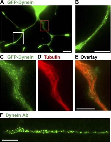
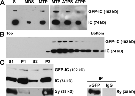
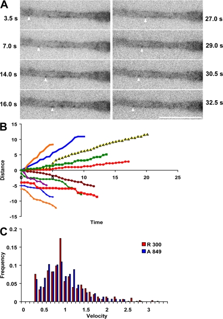
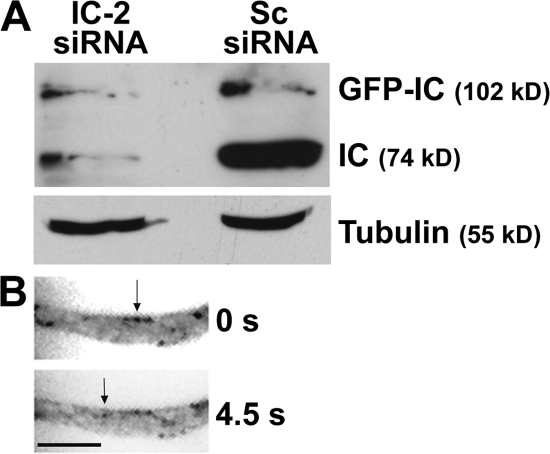
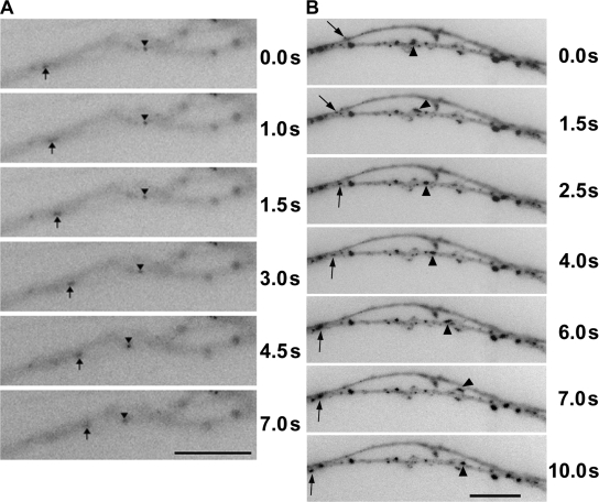
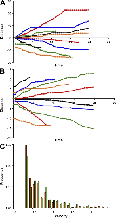
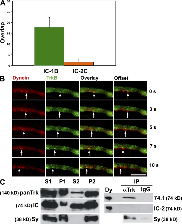
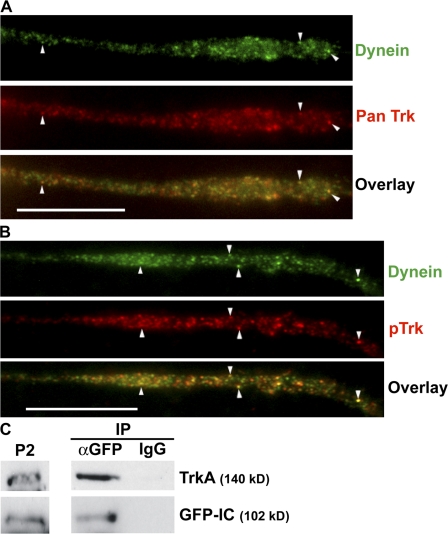
Cytoplasmic dynein is the multisubunit motor protein for retrograde movement of diverse cargoes to microtubule minus ends. Here, we investigate the function of dynein variants, defined by different intermediate chain (IC) isoforms, by expressing fluorescent ICs in neuronal cells. Green fluorescent protein (GFP)-IC incorporates into functional dynein complexes that copurify with membranous organelles. In living PC12 cell neurites, GFP-dynein puncta travel in both the anterograde and retrograde directions. In cultured hippocampal neurons, neurotrophin receptor tyrosine kinase B (TrkB) signaling endosomes are transported by cytoplasmic dynein containing the neuron-specific IC-1B isoform and not by dynein containing the ubiquitous IC-2C isoform. Similarly, organelles containing TrkB isolated from brain by immunoaffinity purification also contain dynein with IC-1 but not IC-2 isoforms. These data demonstrate that the IC isoforms define dynein populations that are selectively recruited to transport distinct cargoes.


Figures

Figure 1.
Figure 1.
Localization of GFP-dynein puncta near microtubules in PC12 cell neurites. (A–E) PC12 cells with stable expression of GFP–IC-2C (GFP-Dynein, green) were differentiated with NGF, fixed, and stained with anti-tubulin (red). (A, B, C, and E) GFP-dynein; (D and E) tubulin. (A) PC12 cell with multiple neurites. (B) Enlargement of a region containing discrete dynein puncta indicated by the white box in A. (C–E) Enlargements of the GFP, tubulin, and overlay images, respectively, of the region indicated by the red box in A. (F) Neurite of an untransfected PC12 cell stained with pan dynein antibody 74.1 (Dynein Ab, green). Bars: (A) 10 μm; (B–F) 5 μm.
Figure 2.
Figure 2.
Biochemical characterization of the incorporation of the GFP–IC-2C isoform into functional cytoplasmic dynein complexes. (A) Microtubule binding. Western blots of the indicated fractions prepared from PC12 cells with stable expression of GFP–IC-2C were probed with a pan antibody to dynein ICs (74.1) that recognized both the endogenous IC (IC) and GFP-tagged IC (GFP–IC). Taxol-stabilized microtubules were added to the soluble fraction (S), and centrifugation yielded the microtubule depleted supernatant (MDS) and microtubule pellet (MTP). The microtubule pellet was resupended in 10 mM Mg-ATP, and centrifugation yielded the ATP supernatant (ATPS) and microtubule pellet (ATPP). The identity of the GFP–IC-2C bands was confirmed with an antibody to GFP (not depicted). Densitometry was used to estimate that the GFP–IC pool was <15% of the total IC pool. (B) Sucrose density gradient sedimentation. The soluble fraction was also fractionated by sucrose density gradient centrifugation, and the proteins in the fractions were analyzed by SDS-PAGE and Western blotting. The blot was probed with the pan dynein antibody. The fractions at the top (5% sucrose) and bottom (20% sucrose) of the gradient are indicated above the blot. (C) Association with membrane-bounded organelles. PC12 cells were lysed in a cytoplasm-like buffer and fractionated; the postnuclear supernatant (S1) and pellet (P1), high-speed supernatant (S2), and high-speed membrane fraction (P2) are indicated. Immunoprecipitation (IP): magnetic beads, preloaded with antibodies to GFP (αGFP) or control IgG (IgG), were incubated with the membrane fraction, P2. The beads were washed and the fractions were analyzed by SDS-PAGE, and the Western blots were probed with the pan dynein antibody and an antibody to synaptophysin (SY), a membranous organelle marker.
Figure 3.
Figure 3.
Bidirectional movement of dynein complexes containing IC-2C in a PC12 cell neurite. (A) Live cell imaging. Individual frames from a video (Video 1, available at http://www.jcb.org/cgi/content/full/jcb.200803150/DC1) of GFP–IC-2C dynein puncta moving in a PC12 cell neurite. (left) Arrowheads indicate GFP–IC-2C dynein puncta moving in the anterograde direction. (right) Arrowheads indicate GFP–IC-2C dynein puncta moving in the retrograde direction. The time from the start of the video is indicated to the right and left of the panels. Bar, 10 μm. (B) Displacement tracking of IC-2C dynein excursions in PC12 cell neurites. The positions of representative individual dynein puncta were tracked along the neurite in each frame of the videos, and the linear displacements (in micrometers) of 11 individual dynein puncta are graphed against time (in seconds) of movement. Anterograde movement is recorded as positive displacement and retrograde movement as negative. The initial positions of the puncta were set to 0, whereas the displacements of some of the retrogradely moving puncta were offset on the y axis to distinguish them on the graph. (C) Interval velocity distribution of dynein containing IC-2C in neurites of the stable PC12 cell line. Velocities (μm/s) of individual movements of GFP–IC-2C dynein puncta between two frames were plotted against the frequency of their occurrence; motility in the anterograde direction (blue) or retrograde direction (red) is indicated. (inset) The number of measurements for the anterograde (A) and retrograde (R) directions.
Figure 4.
Figure 4.
Retrograde movement of dynein complexes containing IC-2C in siRNA-treated PC12 cell neurites. (A) Knockdown of endogenous ICs but not GFP–IC by siRNA. The GFP–IC-2C stable PC12 cell line was transfected with either siRNA oligonucleotides to the 3′ untranslated region of the rat IC-2 gene (IC-2 siRNA) or control siRNA (Sc siRNA) by electroporation. The cells were grown for 72 h after transfection, and cell lysates were analyzed by SDS-PAGE and Western blotting. The blot was probed with the pan IC antibody (74.1) to identify the endogenous ICs (IC) and the GFP–IC-2C (GFP–IC), and, for a loading control, a tubulin antibody (Tubulin). (B) Retrograde dynein movement in siRNA-treated PC12 cell neurites. IC-2 siRNA-transfected PC12 cells with stable expression of GFP–IC-2C were grown on coverslips and differentiated by the addition of NGF. 72 h after transfection, the dynein motility was imaged. The panels show a video of a portion of a PC12 cell neurite (Video 2, available at http://www.jcb.org/cgi/content/full/jcb.200803150/DC1). The arrows indicate one puncta with retrograde movement between the two frames. The time between the video panels is indicated on the right. Although not identified with arrows, many other puncta were moving in the anterograde and retrograde directions. Bar, 5 μm.
Figure 5.
Figure 5.
Bidirectional movement of dynein complexes containing IC-2C and IC-1B in neurons. (A) IC-2C dynein. Individual frames of a video (Video 3, available at http://www.jcb.org/cgi/content/full/jcb.200803150/DC1) showing the movement of dynein puncta in an axon of a cultured hippocampal neuron transfected with mRFP–IC-2C. (B) IC-1B dynein. Individual frames of a video (Video 4) showing movement of dynein puncta in an axon of a cultured hippocampal neuron transfected with mRFP–IC-1B. Arrowheads indicate a puncta moving in the anterograde direction and arrows indicate a puncta moving in the retrograde direction. The time of each frame from the first is indicated on the right. Bars, 10 μm.
Figure 6.
Figure 6.
Comparison of the motility of dynein complexes containing IC-2C and IC-1B in neurons. (A) Displacement tracking of IC-2C dynein. The positions of representative individual dynein puncta were tracked along the neurite in each frame of the videos, and the linear displacements (in micrometers) of 10 individual dynein puncta were graphed against time (in seconds). Anterograde movement is recorded as positive and retrograde movement as negative displacement. (B) Displacement tracking of IC-1B dynein. The positions of representative individual dynein puncta were tracked along the axon in each frame of the videos, and the linear displacements of 10 individual dynein puncta are graphed against time of movement. Anterograde movement is recorded as positive and retrograde movement as negative displacement. The initial positions of many of the puncta were set at 0, whereas the initial displacements of the some puncta were offset on the y axis to distinguish them on the graph. (C) Comparison of the retrograde interval velocity distributions of dynein complexes with the IC-2C and IC-1B isoforms in neurons. Velocities (μm/s) of individual retrograde movements of GFP–IC-2C dynein puncta between two frames were plotted against the frequency of their occurrence. Orange, IC-2C dynein (n = 592); green, IC-1B dynein puncta (n = 692).
Figure 7.
Figure 7.
Coordinate movement of TrkB and cytoplasmic dynein complexes containing IC-1B in neurons. (A) Comparison of the colocalization of TrkB with IC-2C and IC-1B dynein complexes. Hippocampal neurons were cotransfected with TrkB-GFP and either mRFP–IC-2C or mRFP–IC-1B and imaged in living cells; the number of dynein, Trk, and overlapping puncta was then determined in the transfected neurons. The percentages of dynein puncta that colocalized with TrkB were determined for each neuron, averaged, and graphed with the standard error. Dynein containing IC-1B (green) was significantly more likely (P < 0.005, Student's t test) to be associated with the TrkB carrier vesicles than dynein containing IC-2C (orange). 15 IC-1B–transfected neurons were analyzed; a total of 211 dynein puncta and 270 TrkB puncta were counted, and 41 of the puncta were colocalized. 10 IC-2C–transfected neurons were analyzed; a total of 302 dynein puncta and 282 TrkB puncta were counted, and 10 of the puncta were colocalized. (B) Live cell imaging. Hippocampal neurons were cotransfected with TrkB-GFP and mRFP–IC-1B. TrkB-GFP (green) and mRFP-dynein image sets were collected simultaneously as described in the Materials and methods. The panels are individual frames derived from Video 5 (available at http://www.jcb.org/cgi/content/full/jcb.200803150/DC1). The coordinately moving spots are indicated with white arrows. Panels from left to right: (Dynein) movement of the dynein puncta (red); (TrkB) movement of TrkB puncta (green); (Overlay) overlay of the dynein (red) and TrkB (green) puncta, the moving yellow puncta is indicated with an arrow; (Offset) vertical offset of the dynein puncta (red, bottom) and TrkB puncta (green, top) to more clearly demonstrate the coordinate movement of the two proteins. The time interval for each frame from the start of the video is indicated on the right. Bar, 10 μm. (C) IC-1 dynein is preferentially associated with Trk containing vesicles from brain cortex. Membrane-bounded organelles were isolated from fresh rat brain cortex, and the fractions were analyzed by SDS-PAGE and Western blotting; the postnuclear supernatant (S1) and pellet (P1), high-speed supernatant (S2), and high-speed membrane fraction (P2) are indicated. The presence of Trks, ICs, and synaptophysin (Sy) in the fractions was determined by probing the blots with antibodies to Trk and synaptophysin and the pan IC antibody, 74.1. Trk containing organelles were purified by immunoaffinity purification on magnetic beads from the membrane fraction (P2) with antibodies to Trk (αTrk) or control IgG (IgG) and analyzed by SDS-PAGE and Western blotting. The IC isoforms present in the α-Trk immunoprecipitate were identified by probing the blot with an antibody specific to the IC-2 isoforms (IC-2) followed by the pan IC antibody (74.1). As a positive control for the presence of both IC-1 and IC-2 on the blot, dynein immunoprecipitated from the testis was loaded on a neighboring lane (Dy).
Figure 8.
Figure 8.
TrkA signaling endosomes associate with IC-2C dynein complexes in PC12 cells. (A) Colocalization of dynein and the TrkA growth factor receptor in PC12 cells. Neurite of a stable GFP–IC-2C PC12 cell treated with siRNA stained with dynein pan IC antibody, 74.1 (Dynein, green) and a pan Trk antibody (Pan Trk, red), and the overlay of the two colors. Arrowheads indicate some of the colocalizing puncta. (B) Colocalization of dynein and activated TrkA growth factor receptor in PC12 cells. Neurite of stable GFP–IC-2C cell line treated with siRNA stained with dynein pan IC antibody, 74.1 (dynein, green), and an antibody that reacts with activated (phosphorylated) Trk (pTrk, red). Arrowheads indicate some of the colocalizing puncta. Bar, 10 μm. (C) GFP–IC-2C–containing dynein is associated with TrkA containing organelles in PC12 cells. The membrane fraction (P2) was prepared from NGF-stimulated PC12 cells as described for Fig. 2, and GFP–IC–containing vesicles were isolated by immunoaffinity purification on magnetic beads laded with antibodies to GFP. The anti-GFP (α-GFP) and control IgG beads were washed, and the bound proteins were analyzed by SDS-Page and Western blotting with pan Trk antibody and the pan IC antibody, 74.1. The presence of GFP–IC (GFP–IC) on the beads was a positive control.
Figure 9.
Figure 9.
Regulation of dynein by neurotrophins and Trks. (A) Recruitment of activated (phosphorylated) TrkA to dynein containing vesicles in NGF-treated PC12 cells. PC12 cells were harvested and then treated with NGF (+NGF) or buffer (−NGF). The postnuclear supernatant (S1) and the membrane fraction (P2) were prepared and analyzed by SDS-PAGE and Western blotting. The blot was first probed with an antibody specific for the phosphorylated (activated) form of Trk (pTrk). Next, the blot was stripped and probed with pan Trk antibody (panTrk). The vesicle fractions were immunoprecipitated (IP) with anti-pan IC antibody (74.1) or control IgG (IgG) on magnetic beads, and a Western blot of the proteins bound to the beads was probed with the antibody to activated Trk (pTrk). The blots were probed with a pan Trk antibody as a loading control. (B) Modulation of dynein by NGF. (left) Graphs of mean anterograde (blue) and retrograde (red) interval velocities for control (Control) and NGF-treated (+NGF) PC12 cells. The mean interval retrograde velocity for NGF-stimulated cells was 1.2 μm/s ± 0.05 standard error (n = 140), and for control cells 0.9 μm/s ± 0.03 standard error (n = 300). The difference in mean retrograde velocities was significant at P < 0.00017 (Student's t test). (right) Graph showing the percent of GFP–IC-2C dynein puncta moving in the anterograde (blue) and retrograde (red) directions in control (Control) and NGF-stimulated cells (+NGF). The addition of NGF resulted in a significant increase in the relative number of dynein puncta moving in the retrograde direction (P < 0.0002, χ2 test).

References

    1. Arevalo, J.C., D.B. Pereira, H. Yano, K.K. Teng, and M.V. Chao. 2006. Identification of a switch in neurotrophin signaling by selective tyrosine phosphorylation. J. Biol. Chem. 281:1001–1007. - PubMed
    1. Bhattacharyya, A., F.L. Watson, S.L. Pomeroy, Y.Z. Zhang, C.D. Stiles, and R.A. Segal. 2002. High-resolution imaging demonstrates dynein-based vesicular transport of activated Trk receptors. J. Neurobiol. 51:302–312. - PubMed
    1. Bronfman, F.C., M. Tcherpakov, T.M. Jovin, and M. Fainzilber. 2003. Ligand-induced internalization of the p75 neurotrophin receptor: a slow route to the signaling endosome. J. Neurosci. 23:3209–3220. - PMC - PubMed
    1. Campbell, R.E., O. Tour, A.E. Palmer, P.A. Steinbach, G.S. Baird, D.A. Zacharias, and R.Y. Tsien. 2002. A monomeric red fluorescent protein. Proc. Natl. Acad. Sci. USA. 99:7877–7882. - PMC - PubMed
    1. Caviston, J.P., J.L. Ross, S.M. Antony, M. Tokito, and E.L. Holzbaur. 2007. Huntingtin facilitates dynein/dynactin-mediated vesicle transport. Proc. Natl. Acad. Sci. USA. 104:10045–10050. - PMC - PubMed

Publication types

MeSH terms