. 2008 Jun;20(6):1555-66.
doi: 10.1105/tpc.108.058578. Epub 2008 Jun 17.

Arabidopsis ELONGATED MITOCHONDRIA1 is required for localization of DYNAMIN-RELATED PROTEIN3A to mitochondrial fission sites

Affiliations

Arabidopsis ELONGATED MITOCHONDRIA1 is required for localization of DYNAMIN-RELATED PROTEIN3A to mitochondrial fission sites

Shin-ichi Arimura et al. Plant Cell. 2008 Jun.

Abstract

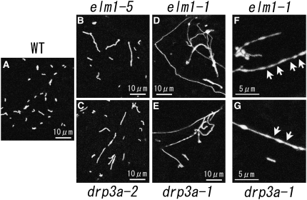
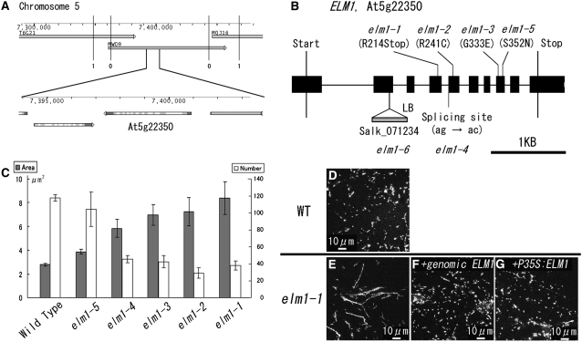
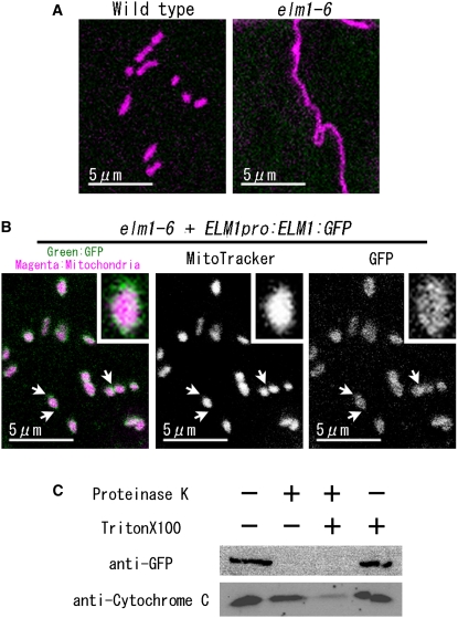
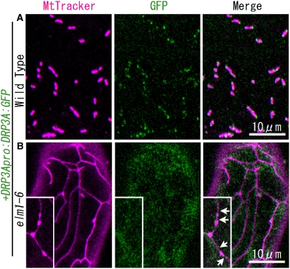
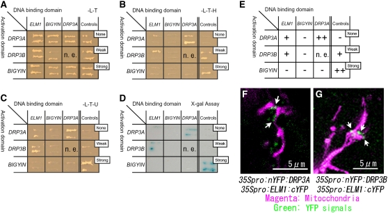
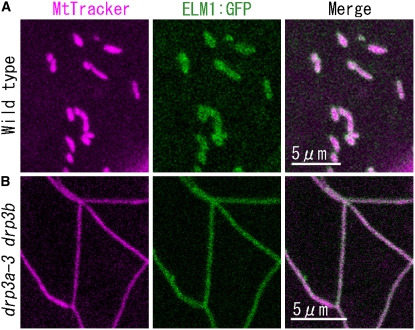
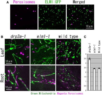
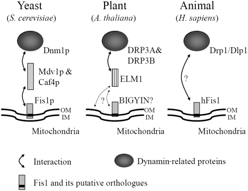
Mitochondrial fission is achieved partially by the activity of self-assembling dynamin-related proteins (DRPs) in diverse organisms. Mitochondrial fission in Arabidopsis thaliana is mediated by DRP3A and DRP3B, but the other genes and molecular mechanisms involved have yet to be elucidated. To identify these genes, we screened and analyzed Arabidopsis mutants with longer and fewer mitochondria than those of the wild type. ELM1 was found to be responsible for the phenotype of elongated mitochondria. This phenotype was also observed in drp3a plants. EST and genomic sequences similar to ELM1 were found in seed plants but not in other eukaryotes. ELM1:green fluorescent protein (GFP) was found to surround mitochondria, and ELM1 interacts with both DPR3A and DRP3B. In the elm1 mutant, DRP3A:GFP was observed in the cytosol, whereas in wild-type Arabidopsis, DRP3A:GFP localized to the ends and constricted sites of mitochondria. These results collectively suggest that mitochondrial fission in Arabidopsis is mediated by the plant-specific factor ELM1, which is required for the relocalization of DRP3A (and possibly also DRP3B) from the cytosol to mitochondrial fission sites.


Figures

Figure 1.
Figure 1.
Mitochondrial Morphologies in drp3a and elm1 Mutants. Mitochondria in leaf epidermal cells of wild-type plants (A) and in weak (elm1-5 [B] and drp3a-2 [C]) and severe (elm1-1 [D] and ]F] and drp3a-1 [E] and [G]) mutants transformed with Mt-GFP. Arrows indicate the constricted sites of elongated mitochondria.
Figure 2.
Figure 2.
ELM1 Is the Gene Responsible for the Elongation of Mitochondria in elm1 Mutants. (A) Map-based cloning of ELM1. The numbers of recombination events between the molecular markers and the ELM1 locus are shown under the vertical bars (0 and 1). T6G21, MWD9, and MQJ16 are overlapping BAC clones. Numbers on the gauges show the physical distance in base pairs from the northern end of chromosome 5. The figure was modified from the Arabidopsis Genome Initiative Map of Map Viewer in The Arabidopsis Information Resource home page. (B) The positions of base pair substitutions in elm1-1 through elm1-5 mutants and the DNA insertion in elm1-6. ELM1 consists of 10 exons (black boxes) and nine introns (black lines). (C) Mitochondrial sizes and numbers in epidermal cells of wild-type and elm1 mutants. Gray bars show the average planar area (in μm2) of the mitochondria (n > 90). White bars show the average numbers of mitochondria in a 120 μm × 120 μm area (n = 3). All error bars show se. (D) to (G) Complementation test of mitochondrial elongation in elm1-1. The wild type (D), elm1-1 (E), and elm1-1 (F) transformed with a genomic fragment of the ELM1 gene with 950 bp upstream region of the start codon to 345 bp downstream of the stop codon (genomic ELM1) and elm1-1 (G) with P35S:ELM1-ORF. Both restored mitochondrial morphology to that of the wild type.
Figure 3.
Figure 3.
ELM1:GFP Localizes to the Outer Surface of Mitochondria. (A) and (B) ELM1:GFP rescued the elongation of mitochondria in elm1-6 and localized on mitochondria. Mitochondria in epidermal cells of the cotyledon were visualized as magenta by MitoTracker Orange CMTMRos staining. (B) A genomic fragment of ELM1 fused to GFP (ELM1pro:ELM1:GFP) was introduced into elm1-6. Insets show 4× magnified images of mitochondria. Arrows show constricted sites and end sites of mitochondria where green signals seem more intense. (C) Protease protection assay. Isolated mitochondria from elm1-6 transformed with ELM1pro:ELM1:GFP were incubated in the presence or absence of Proteinase K or Triton X-100, as indicated. The samples were subjected to SDS-PAGE, and protein gel blotting was done with anti-GFP and anti-rice cytochrome C antiserum. A representative result of three technical repeats and two biological repeats is shown.
Figure 4.
Figure 4.
Plant Growth of the elm1-1 Mutant. Wild-type and elm1-1 plants on the 13 d (A) and 26 d (B) after germination. The wild type is a Mt-GFP transgenic line. Bars = 3 cm.
Figure 5.
Figure 5.
DRP3A:GFP Does Not Localize to Mitochondrial Ends and Constricted Sites in the elm1-6 Mutant. Confocal laser scanning microscopy images of the cotyledon leaf epidermal cells of wild-type plants (A) and elm1-6 T-DNA insertion mutants (B) transformed with the construct DRP3Apro:DRP3A:GFP. Mitochondria were visualized by MitoTracker Orange staining. Insets in (B) show beads-on-a-string-shaped mitochondria that transiently appeared in elm1-6 from a separate microscope field. Arrows show the constricted sites on mitochondria in elm1-6. Bar in the bottom panel indicates scale of insets.
Figure 6.
Figure 6.
ELM1 Interacts with DRP3A and DRP3B. (A) to (C) Y2H analysis. Yeast strain Mav203 was transformed with paired constructs of protein fused to the GAL4 activation domain (left) or the GAL4 DNA binding domain (top). Controls show the combinations with no, weak, and strong interactions, as indicated (see Methods). Transformants were streaked onto a SD/-Leu/-Thr plate (A), confirming the introduction of both plasmids into the cells; a SD/-Leu/-Thr/-His plate (B), determining the interaction between fusion proteins; and a SD/-Leu/-Thr/-Ura plate (C), determining the interaction between fusion proteins by Ura synthase gene expression. (D) X-Gal assay to check the interaction of fusion proteins by β-galactosidase expression. (E) Summary of the Y2H analysis from (A) to (D). –, no signal; +, weak signal; ++, strong signal; n.e., not examined. (F) and (G) Fluorescence images of Arabidopsis leaf epidermal cells transiently transformed with BiFC formation constructs as indicated, together with a construct for expressing mitochondrial-targeted DsRed by particle bombardment. nYFP, N-terminal half of YFP; cYFP, C-terminal half of YFP. If the two parts of YFP (nYFP and cYFP) come together by interactions between each fused protein (DRP3A and ELM1 in [F]; DRP3B and ELM1 in [G]), they give off a yellow–green fluorescence on excitation. We used a GFP filter set, which made the fluorescence appear green. Arrows show the green spots that indicate protein interaction on mitochondria.
Figure 7.
Figure 7.
ELM1:GFP Localizes on Mitochondria in Both the Wild Type and the drp3a-3 drp3b Mutant. Confocal laser scanning microscopy images of the cotyledon leaf epidermal cells of the wild type (A) and a drp3a-3 drp3b double T-DNA insertion homoline (B) introduced with the construct ELM1pro:ELM1:GFP and stained with MitoTracker Orange.
Figure 8.
Figure 8.
Induction of ELM1 Gene Expression in the elm1-6 Mutant Relocates DRP3A:GFP from the Cytosol to Mitochondria and Restores Wild-Type Mitochondrial Morphology. (A) to (D) Representative images of mitochondria (magenta, stained by MitoTracker Orange) and DRP3A:GFP (green) in elm1-6 transformed with pER8:ELM1 and DRP3A:GFP before (A) and after ([B] to [D]) addition of 17-β estradiol. Bars = 5 μm. (E) The percentage of transformed cells in each phase of mitochondrial fission before and after addition of 17-β estradiol (estr). Phase I (A) is characterized by cells with elongated mitochondria and cytosolic DRP3A:GFP. In phase II, DRP3A:GFP relocalizes to the elongated mitochondria (shown in [B] and [C]). In phase III, DRP3A:GFP is found on the ends of mitochondrial particles (D). Average percentages of cells in each phase for each time point are shown by bars, and lines indicate sd. More than 50 cells were counted each time in triplicate.
Figure 9.
Figure 9.
Loss of ELM1 Does Not Change Peroxisome Morphology. (A) Confocal laser scanning image of the leaf epidermis of a transgenic plant with ELM1pro:ELM1:GFP and peroxisomal-localized DsRed. (B) Mitochondria and peroxisomes in leaf epidermis and root epidermis in drp3a-1, elm1-1, and the wild type were highlighted by GFP (mitochondria) and DsRed (peroxisomes). (C) The average planar area of peroxisomes in leaf epidermis in drp3a-1, elm1-1, and wild-type plants (n > 60). Error bars show se.
Figure 10.
Figure 10.
Comparison of Mitochondrial Fission Factors in Yeast, a Plant, and an Animal. In yeast, a dynamin-related protein, Dnm1p, which is a mitochondrial fission executor, is recruited from the cytosol to mitochondria in a manner dependent on Fis1p and an adaptor protein, Mdv1p (and a Mdv1p-homolog, Caf4p). In the plant Arabidopsis, dynamin-related proteins, DRP3A and DRP3B, interact with ELM1 for their localization to mitochondria. Although ELM1 does not have sequence similarity to Mdv1p or Caf4p, its role may be similar to the roles of Mdv1p and Caf4p. In human, localization of dynamin-related protein, Drp1/Dlp1, to mitochondria depends on hFis1. Other proteins, such as Mdv1p, Caf4p, and ELM1, may also be involved. A black bar in Fis1 shows putative transmembrane domains. OM, outer membrane; IM, inner membrane.

References

    1. Arimura, S., Aida, G.P., Fujimoto, M., Nakazono, M., and Tsutsumi, N. (2004. a). Arabidopsis dynamin-like protein 2a (ADL2a), like ADL2b, is involved in plant mitochondrial division. Plant Cell Physiol. 45 236–242. - PubMed
    1. Arimura, S., and Tsutsumi, N. (2002). A dynamin-like protein (ADL2b), rather than FtsZ, is involved in Arabidopsis mitochondrial division. Proc. Natl. Acad. Sci. USA 99 5727–5731. - PMC - PubMed
    1. Arimura, S., Yamamoto, J., Aida, G.P., Nakazono, M., and Tsutsumi, N. (2004. b). Frequent fusion and fission of plant mitochondria with unequal nucleoid distribution. Proc. Natl. Acad. Sci. USA 101 7805–7808. - PMC - PubMed
    1. Bell, C.J., and Ecker, J.R. (1994). Assignment of 30 microsatellite loci to the linkage map of Arabidopsis. Genomics 19 137–144. - PubMed
    1. Bhar, D., Karren, M.A., Babst, M., and Shaw, J.M. (2006). Dimeric Dnm1-G385D interacts with mdv1 on mitochondria and can be stimulated to assemble into fission complexes containing Mdv1 and Fis1. J. Biol. Chem. 281 17312–17320. - PubMed

Publication types

MeSH terms

Substances