

Assessment of benign tumor burden by whole-body MRI in patients with neurofibromatosis 1
- PMID: 18559970
- PMCID: PMC2666233
- DOI: 10.1215/15228517-2008-011
Assessment of benign tumor burden by whole-body MRI in patients with neurofibromatosis 1
Abstract
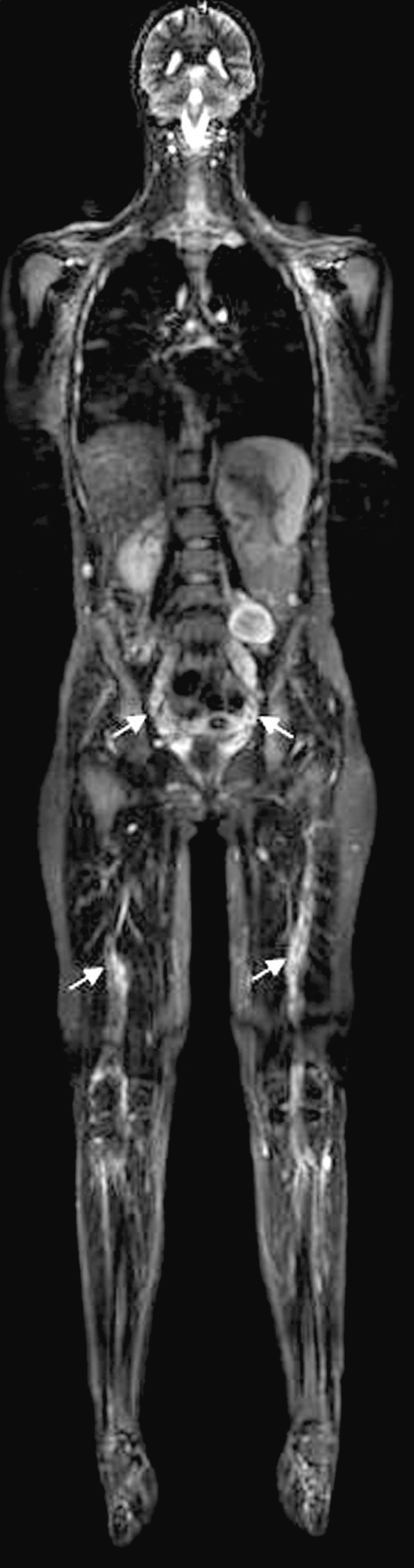
People with neurofibromatosis 1 (NF1) have multiple benign neurofibromas and a 10% lifetime risk of developing malignant peripheral nerve sheath tumors (MPNSTs). Most MPNSTs develop from benign plexiform neurofibromas, so the burden of benign tumors may be a risk factor for developing MPNST. We studied 13 NF1 patients with MPNSTs and 26 age- and sex-matched controls (NF1 patients who did not have MPNSTs) with detailed clinical examinations and whole-body MRI to characterize their body burden of internal benign neurofibromas. Internal plexiform neurofibromas were identified in 22 (56%) of the 39 NF1 patients studied. All six of the NF1 patients with MPNSTs under 30 years of age had neurofibromas visualized on whole-body MRI, compared to only 3 of 11 matched NF1 controls under age 30 (p < 0.05). Both the median number of plexiform neurofibromas (p < 0.05) and the median neurofibroma volume (p < 0.01) on whole-body MRI were significantly greater among MPNST patients younger than 30 years of age than among controls. No significant differences in whole-body MRI findings were observed between NF1 patients with MPNSTs and controls who were 30 years of age or older. Whole-body MRI of NF1 patients allows assessment of the burden of internal neurofibromas, most of which are not apparent on physical examination. Whole-body imaging of young NF1 patients may allow those at highest risk for developing MPNST to be identified early in life. Close surveillance of these high-risk patients may permit earlier diagnosis and more effective treatment of MPNSTs that develop.
Figures
References
-
- Korf B. Clinical features and pathobiology of neurofibromatosis 1. J Child Neurol. 2002;17:573–577. 602–604, 646–651. - PubMed
-
- Thomson S, Fishbein L, Wallace M. NF1 mutations and molecular testing. J Child Neurol. 2002;17:555–561. 571–572, 646–651. - PubMed
-
- Viskochil D. Genetics of neurofibromatosis 1 and the NF1 gene. J Child Neurol. 2002;17:562–572. 646–651. - PubMed
-
- Friedman JM, Riccardi VM. Clinical and epidemiological features. In: Friedman JM, Gutmann DH, MacCollin M, Riccardi VM, editors. Neurofibromatosis: Phenotype, Natural History, and Pathogenesis. 3rd ed. Baltimore, MD: Johns Hopkins University Press; 1999. pp. 29–86.
-
- Riccardi V. The genetic predisposition to and histogenesis of neurofibromas and neurofibrosarcoma in neurofibromatosis type 1. Neurosurg Focus. 2007;22(6):E3. - PubMed
MeSH terms
LinkOut - more resources
Full Text Sources
Medical
Research Materials
Miscellaneous
