. 2008 Sep 1;112(5):1960-70.
doi: 10.1182/blood-2007-09-113860. Epub 2008 Jun 17.

Absence of SKP2 expression attenuates BCR-ABL-induced myeloproliferative disease

Affiliations

Absence of SKP2 expression attenuates BCR-ABL-induced myeloproliferative disease

Anupriya Agarwal et al. Blood. .

Abstract

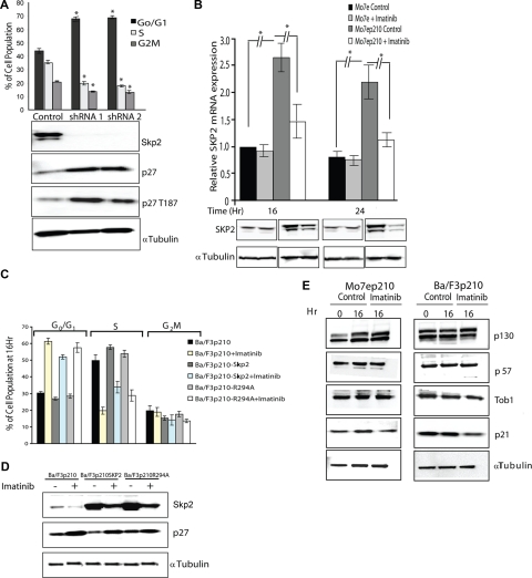
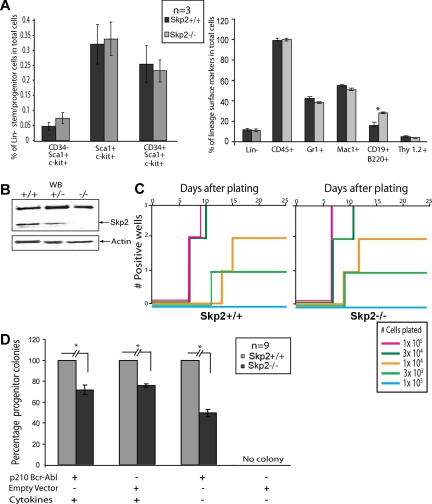
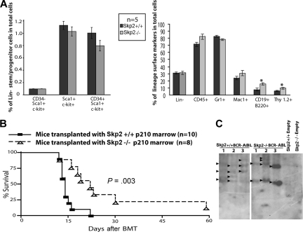
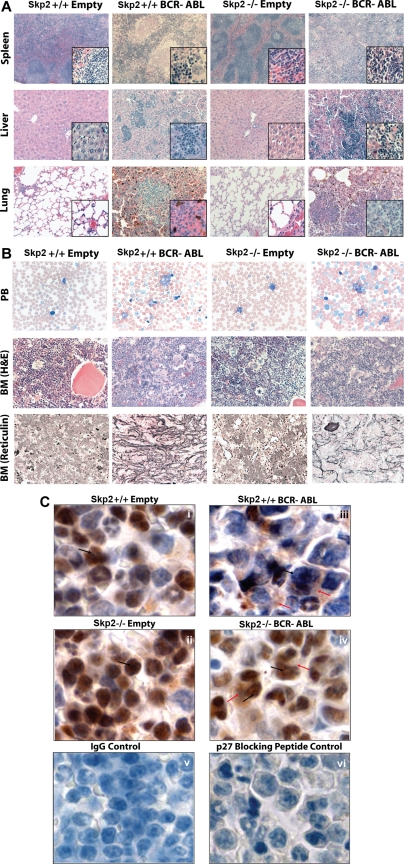
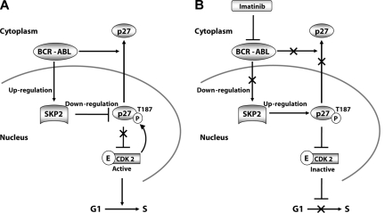
BCR-ABL is proposed to impair cell-cycle control by disabling p27, a tumor suppressor that inhibits cyclin-dependent kinases. We show that in cell lines p27 expression is inversely correlated with expression of SKP2, the F-box protein of SCF(SKP2) (SKP1/Cul1/F-box), the E3 ubiquitin ligase that promotes proteasomal degradation of p27. Inhibition of BCR-ABL kinase causes G(1) arrest, down-regulation of SKP2, and accumulation of p27. Ectopic expression of wild-type SKP2, but not a mutant unable to recognize p27, partially rescues cell-cycle progression. A similar regulation pattern is seen in cell lines transformed by FLT3-ITD, JAK2(V617F), and TEL-PDGFRbeta, suggesting that the SKP2/p27 conduit may be a universal target for leukemogenic tyrosine kinases. Mice that received transplants of BCR-ABL-infected SKP2(-/-) marrow developed a myeloproliferative syndrome but survival was significantly prolonged compared with recipients of BCR-ABL-expressing SKP2(+/+) marrow. SKP2(-/-) leukemic cells demonstrated higher levels of nuclear p27 than SKP2(+/+) counterparts, suggesting that the attenuation of leukemogenesis depends on increased p27 expression. Our data identify SKP2 as a crucial mediator of BCR-ABL-induced leukemogenesis and provide the first in vivo evidence that SKP2 promotes oncogenesis. Hence, stabilization of p27 by inhibiting its recognition by SCF(SKP2) may be therapeutically useful.


Figures

Figure 1
Figure 1
Inhibiting BCR-ABL kinase activity up-regulates p27 and down-regulates SKP2. Mo7e cells expressing p210BCR-ABL were treated with imatinib (2.5 μM) for 16 hours. (A) Immunoblot analysis of whole-cell lysates with antiphosphotyrosine antibodies. (B) The cell-cycle profile was analyzed by propidium iodide (PI) staining. Mo7e cells treated with imatinib (2.5 μM) for 16 hours were used as a control. (C) Immunoblot analysis of the effect of BCR-ABL kinase inhibition on p27, cyclin E, CDK2, pT187p27, SKP2, CKS1, KPC1, KPC2 expression levels in nuclear (N) and cytoplasmic extracts (C). Sp1 and α-tubulin were used to assess the purity of the nuclear and cytoplasmic fractions, respectively, and actin was used as a loading control. No effect on p27 and SKP2 expression was observed in Mo7e cells treated with imatinib under similar conditions. (D) Analysis of p27 in nuclear (N) and cytoplasmic (C) lysates of Mo7e p210BCR-ABL cells shows accumulation of p27 predominantly in the nucleus compared with the cytoplasm. Densitometry was performed to quantitate p27 levels from 3 independent experiments. All densitometry values are given in Boehringer light units (BLU). (E) Inhibition of BCR-ABL kinase activity with imatinib reduces CDK2 activity toward histone H1. Mo7e cells expressing p210BCR-ABL were treated with 2.5 μM imatinib for 16 hours and lysates from whole cells as well as from nuclear and cytoplasmic fractions were immunoprecipitated with an antibody against CDK2. Immunoprecipitates were incubated in kinase buffer in the presence of [γ32P]-ATP and histone H1 as a substrate. CDK2 kinase activity toward histone H1 was measured by scintillation counting in 3 independent experiments. Error bars represent SD.
Figure 2
Figure 2
p27 levels are regulated by SKP2 in BCR-ABL-positive cell lines. (A) Mo7ep210BCR-ABL cells were infected with lentivirus-producing SKP2 shRNA-1 and -2 or control (scramble) shRNA construct for stable knockdown of SKP2. The effect on cell-cycle distribution (PI staining) as well as on SKP2, p27, and pT187p27 expression was analyzed by immunoblot analysis. The differences in cell-cycle progression after knockdown of SKP2 are significant compared with control with P values less than .001 from 3 independent experiments. (B) SKP2 mRNA levels were assessed in Mo7e and Mo7ep210BCR-ABL cells by quantitative RT-PCR after treatment with 2.5 μM imatinib for 16 and 24 hours. (C) Ba/F3 cells expressing p210BCR-ABL were stably transduced with a Skp2 expression vector and treated with 2.5 μM imatinib for 16 hours. Cell-cycle distribution was analyzed by PI staining in 3 independent experiments, and (D) p27 expression was analyzed by immunoblot. (E) Effect of BCR-ABL kinase inhibition on the expression of SKP2 substrates was analyzed. Cells were treated with 2.5 μM imatinib and the expression of p130, p57, Tob1, and p21 was measured by immunoblot analysis in Mo7ep210BCR-ABL and Ba/F3p210BCR-ABL cells. Error bars represent SE.
Figure 3
Figure 3
Effect of growth factor and serum deprivation or stimulation on BCR-ABL–positive cells. Parental and BCR-ABL–positive cell lines were deprived or stimulated with growth factor and/or serum for 16 hours. Effect on cell-cycle progression (PI staining) as well as p27 and Skp2 expression was analyzed in 3 independent experiments. BCR-ABL–positive cells were also treated with 2.5 μM imatinib. (A) Mo7e and Mo7ep210BCR-ABL cells. (B) Ba/F3 and Ba/F3p210BCR-ABL cells. Error bars represent SE. (C) Effect on Skp2 and p27 expression of inhibiting constitutively active tyrosine kinases in leukemia cell lines. Cells were incubated with the indicated concentrations of inhibitors for 24 hours and immunoblot analysis was performed on whole cell lysates. Left panel shows MOLM14 cells expressing a FLT3 internal tandem duplication (ITD) treated with 500 nM MLN518. Middle panel shows HEL cells expressing JAK2 V617F treated with 50, 75, 100 μM of AG490. Right panel shows Ba/F3 cells expressing TEL-PDGFRβ treated with 2.5, 5.0, 10 μM imatinib. Skp2 and p27 expression levels were analyzed after inhibition of tyrosine kinase activity. α-Tubulin was used as a loading control.
Figure 4
Figure 4
In vitro transformation of primary bone marrow cells by BCR-ABL in Skp2+/+ versus Skp2−/− bone marrow. (A) Flow cytometric analysis of non–5-FU-treated Skp2+/+ and Skp2−/− mice bone marrow cells for stem/multipotent progenitor population (left) and lineage surface markers (right). Significant differences (P < .05) are indicated by an asterisk. (B) Immunoblot showing Skp2 expression from bone marrow cells of Skp2+/+, Skp2+/−, and Skp2−/− littermates. Actin was used as a loading control. (C) Comparison of B-cell transformation by BCR-ABL between Skp2+/+ and Skp2−/− bone marrow cells. C57/BL6 bone marrow cells were transduced with p210BCR-ABL or empty vector retroviral supernatant and the indicated numbers of transduced viable cells were plated in Whitlock-Witte culture medium in triplicate on stromal cells (106 cells/well) derived from untransduced marrow. Wells were scored as positive when the numbers of viable nonadherent cells reached 106/well. No growth was observed in Skp2+/+ or Skp2−/− bone marrow cells transduced with empty vector retrovirus. Data shown here are representative of 3 independent experiments. (D) Comparison of myeloid colony formation between Skp2+/+ and Skp2−/− bone marrow cells. Bone marrow cells were transduced with p210BCR-ABL or empty vector retrovirus and plated in methylcellulose in the presence or absence of cytokines. The histogram shows the average percentage of remaining colonies for Skp2−/− marrow cells (considering Skp2+/+ as 100%) from triplicate assays from 3 independent experiments. The difference between Skp2+/+ and Skp2−/− mice is significant (P < .001). No colonies were recovered from cells transduced with empty vector and grown in the absence of cytokines. Mice used for in vitro assays are not treated with 5-FU. Error bars represent SE.
Figure 5
Figure 5
In vivo evidence for decreased leukemogenicity in Skp2−/− mice. (A) Flow cytometric analysis of 5-FU–treated Skp2+/+ and Skp2−/− mice bone marrow cells for stem/multipotent progenitor population (left) and lineage surface markers (right). Significant differences (P < .05) are indicated by an asterisk. Error bars represent SE. (B) Kaplan-Meier survival curve for mice transplanted with Skp2+/+ and Skp2−/− marrow transduced with p210BCR-ABL. The number of individual mice in each arm is indicated. Mice transplanted with Skp2−/− marrow survived significantly longer (P = .003). (C) Southern blot analysis for proviral integration from BCR-ABL or empty vector transduced Skp2+/+ and Skp2−/− mice. Genomic DNA from spleen tissue (15 μg) was digested with restriction enzyme BglII, resolved on a 1.5% agarose gel, and transferred to Hybond-N+ membrane. The blots were hybridized with a 32P-labeled GFP probe and exposed to autoradiography film.
Figure 6
Figure 6
Representative histology of mice transplanted with Skp2+/+ and Skp2−/− marrow transduced with p210BCR-ABL or empty vector. (A) Spleen, liver, and lung [hematoxylin and eosin (H&E) stain, original magnifications are ×5, ×20, and ×20, respectively]. Higher magnification ×40 is also shown in the lower right corners. (B) Peripheral blood smear (Wright-Giemsa stain, original magnification ×63), and bone marrow (H&E stain and reticulin fiber stain, original magnification ×40). (C) p27 Immunohistochemistry of spleen of mice that received Skp2+/+ and Skp2−/− marrow transduced with empty vector (i, ii) or p210BCR-ABL (iii, iv). p27 Staining was performed using polyclonal anti-p27 primary antibody (C19; Santa Cruz Biotechnology) and biotinylated anti–rabbit IgG (H&L) secondary antibody (Vector Laboratories, Burlingame, CA) using Vectastain Elite Universal ABC Kit (Vector Laboratories) and DAB Chromogen (Dako, Carpinteria CA). Nuclear and cytoplasmic p27 are indicated by black and red arrows, respectively. For negative controls, staining with IgG antibody (v) and with anti-p27 antibody including p27-specific blocking peptide (Santa Cruz Biotechnology) showing practically complete block of p27 staining in cells with some residual background staining (vi). Original magnifications, ×100. Images were viewed with a Leica DM LB2 microscope (Leica, Wetzlar, Germany) equipped with Leica N Plan 5×/0.12 (for 5× magnification), HC PL Fluotar 20×/0.50 (20×), HCX PL Fluotar 40×/0.75 (40×), C Plan 63×/0.75 (63×), and N Plan 100×/1.25 oil (100×) objective. Images were captured with a Leica DC300 camera running IM50 Image Manager software (v5 release 222; Heerbrugg, Switzerland) and processed with Adobe Photoshop CS2 9.0.2 (Adobe Systems, San Jose, CA).
Figure 7
Figure 7
Schematic representation of SKP2-mediated regulation of p27 in BCR-ABL–positive cell lines. (A) BCR-ABL kinase activity induces expression of SKP2, which increases the activity of SCFSKP2, which in turn promotes p27 degradation. This releases CDK2 from p27 inhibition and stimulates cell-cycle progression in BCR-ABL positive cell lines. In primary cells, BCR-ABL additionally promotes cytoplasmic localization of p27. (B) Inhibition of BCR-ABL kinase activity by imatinib decreases SKP2 expression, which leads to accumulation of pT187p27, which restores inhibition of CDK2.

Similar articles

Cited by

References

    1. Deininger MW, Goldman JM, Melo JV. The molecular biology of chronic myeloid leukemia. Blood. 2000;96:3343–3356. - PubMed
    1. Clarkson B, Strife A, Wisniewski D, Lambek CL, Liu C. Chronic myelogenous leukemia as a paradigm of early cancer and possible curative strategies. Leukemia. 2003;17:1211–1262. - PubMed
    1. Attar EC, Scadden DT. Regulation of hematopoietic stem cell growth. Leukemia. 2004;18:1760–1768. - PubMed
    1. Skorski T, Kanakaraj P, Nieborowska-Skorska M, et al. Phosphatidylinositol-3 kinase activity is regulated by BCR/ABL and is required for the growth of Philadelphia chromosome-positive cells. Blood. 1995;86:726–736. - PubMed
    1. Cortez D, Reuther G, Pendergast AM. The Bcr-Abl tyrosine kinase activates mitogenic signaling pathways and stimulates G1-to-S phase transition in hematopoietic cells. Oncogene. 1997;15:2333–2342. - PubMed

Publication types

MeSH terms

Substances