. 2008 Sep 1;112(5):1720-9.
doi: 10.1182/blood-2007-09-112748. Epub 2008 Jun 17.

Overexpression of delta-like 4 induces arterialization and attenuates vessel formation in developing mouse embryos

Affiliations

Overexpression of delta-like 4 induces arterialization and attenuates vessel formation in developing mouse embryos

Alexandre Trindade et al. Blood. .

Abstract

The importance of Notch signaling pathway in the regulation of vascular development and angiogenesis is suggested by the expression of Notch receptors and ligands in vascular endothelial cells (ECs) and the observed vascular phenotypes in mutants of Notch receptors or ligands, especially Dll4. DLL4 is specifically expressed in arterial ECs during development, and haplo-insufficiency is embryonically lethal in mice. To address the role of Dll4 in vascular development, we produced mDll4 conditionally overexpressed transgenic mice that were crossed with constitutive recombinase cre lines. Double transgenic embryos displayed grossly enlarged dorsal aortae (DA) and died before embryonic day 10.5 (E10.5), showing a variable degree of premature arteriovenous fusion. Veins displayed ectopic expression of arterial markers. Other defects included reduced vascular sprouting, EC proliferation, and migration. mDll4 overexpression also inhibited VEGF signaling and increased fibronectin accumulation around the vessels. In vitro and in vivo studies of DLL4-FL (Dll4-full-length) in ECs recapitulate many of the mDll4 transgenics findings, including decreased tube formation, reduced vascular branching, fewer vessels, increased pericyte recruitment, and increased fibronectin expression. These results establish the role of Dll4 in arterial identity determination, and regulation of angiogenesis subject to dose and location.


Figures
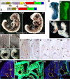
Figure 1
Figure 1
mDll4 overexpression causes major defects in the developing vascular system. (A) pZ/EG-mDll4 transgenesis vector and result of Cre recombination. (B) LacZ staining of a ZEG-Dll4 embryo at embryonic day 8.0 (E8.0). (C) EGFP expression in the double transgenic (DT) embryos at E8.5. (D) Haemorrhaging in the DT embryos at E9.0. Whole-mount PECAM1 immunostaining of E9.0 DT (E) and control (F) embryos. (F) DT embryo at E9.0 showing a hypertrophied dorsal aorta (red arrow), ramified ACV (green arrow), and an unbranched vascular plexus in the head region (blue arrow), relative to the control embryo (E). PECAM1 immunostaining of vitelline membranes from WT (G) and DT embryos (H) show a blockage in the angiogenic remodeling of the vitelline vasculature of mutant embryos. (I,J) Serial sections of an E9.5 WT embryo demonstrating the region where the ACV (green arrow) connects to the sinus venosus (blue arrow; K-M) Serial sections of a E9.5 DT embryo (anterior-posterior) showing a fusion between the aorta (red arrow) and the ACV (green arrow) just before its connection to the sinus venosus (blue arrow). In panel K the ACV consists of a plexus of small capillaries (green arrow) that join to form a single vessel with a large lumen just before its fusion with the dorsal aorta (M). Microangiography with India Ink injection confirmed the existence of functional connections between the DA and the ACV of DT embryos (O), with ink flowing directly from the aortae (red arrow) to the sinus venosus (blue arrow), in contrast to the regular flow of the control embryos (N). Endothelial-specific overexpression of mDll4 (Dll4e) causes the same vascular defects as observed in DT embryos. (P) Immunofluorescence with anti-PECAM1 and anti-Dll4 antibodies on WT embryo at E9.5. Immunofluorescence with anti-PECAM1 and anti-Dll4 antibodies on DT embryo (Q,R) and Dll4e embryos at E9.5 (S,T), confirming that Dll4 is ubiquitously expressed in DT embryos and that it is pan-endothelial in Dll4e embryos, while WT embryos show only Dll4 expression on the dorsal aortae (DA).
Figure 2
Figure 2
mDll4 overexpression causes morphogenetic defects in the developing heart. PECAM1 immunostaining of cryosections from E9.5 WT and DT embryos (A) WT heart ventricle (v) showing normal trabeculation. (B) DT heart ventricle (v) revealing reduced trabeculation. (C) WT heart atria (a). (D) DT heart atria (a) revealing gross hypertrophy. (E) Outflow tract of heart from WT embryo. (F) Outflow tract of heart from DT embryo, showing acellularization of the outflow tract cushions (*). SMA immunostaining of cryosections from E9.5 WT and DT embryos (G) heart ventricle (v) of WT embryo. (H) Heart ventricle (v) of DT embryo showing aggregation of cardiomyocites at the periphery of the ventricle and irregular distribution of cardiomyocites through the trabeculations, relative to WT. Whole-mount PECAM1 immunostaining of E9.0 WT and DT embryos. (I) WT embryo imaged at the heart level, showing normal arteries at the first (green arrow) and second branchial arches (blue arrow; DA, red arrow). (J) DT embryo imaged at the heart level, showing the presence of a fully formed artery at the first branchial arch (blue arrow) but only a very reduced or nonexistent artery at the second branchial arch (green arrow; DA, red arrow).
Figure 3
Figure 3
Arterial markers are expressed in all endothelial cells while venous markers are absent in E9.0 DT embryos in situ hybridization. (A,D) hey1 mRNA, (B,E) notch1 mRNA. The Notch markers tested show concomitant expression in the aortae (DA) and ACV of DT embryos. Control embryos (D,E) only show expression of these genes in the endothelium of the aortae. (C,F) flk1 mRNA shows lowered expression in the DT embryos. (G,J) ephrin-b2 mRNA, (H,K) connexin37 mRNA. The arterial endothelial specific markers tested show concomitant expression in the aortae and ACV of DT embryos. Control embryos (J,K) only show expression of these genes in the endothelium of the aortae. (I,L) eph-B4 mRNA is not detectable in the ACV of DT embryos despite being detected in the ACV of control embryos (L).
Figure 4
Figure 4
Proliferation of arterial ECs is decreased in DT embryos. (A) BrdU incorporation studies show 16% of ECs per aortic cross-section of DT embryo are proliferating, compared with 28% in WT embryos (P < .01). Number of ECs per aortic cross-section is increased in DT embryos. (B) In WT embryos there are on average 8.5 ECs per aortic cross-section, in DT embryos there are 11.5 ECs, representing a 35% increase (P < .01). Apoptosis of arterial ECs is increased in DT embryos. (C) In WT embryos, on average, 2.6% of arterial ECs are apoptotic, in DT embryos, on average, that frequency is 4.7%, representing a 80% increase in arterial endothelial apoptosis frequency (P < .01). Endothelial cell migration is delayed in DT embryos. Wholemount PECAM1 immunostaining of E8.5 and E9.5 WT and DT embryos. (D) WT embryo at E8.5. (E) DT embryo at E8.5 showing a delay in the migration of ECs from the DA to form the intersomitic blood vessels, relative to control embryo (D). (F) WT embryo at E8.5. (G) DT embryo at E9.5 showing a continued delay in the migration of ECs from the DA to form the intersomitic blood vessels, relative to control embryo (F). Angiogenic sprouting is reduced in DT embryos (H) Cephalic region of WT embryo at E9.5. (I) DT embryo at E9.5 showing a lowered number of new blood vessel sprouts in cephalic region, relative to control embryo.
Figure 5
Figure 5
Extracellular matrix deposition around the dorsal aortae is increased in mDll4 overexpressing embryos: Immunofluorescence of cryosections from E8.5 DT and control embryos. (A,C,D) Fibronectin deposition around the dorsal aorta of a WT embryo. (B,E,F) DT embryo section showing increased amount of fibronectin surrounding the dorsal aorta, relative to control embryo. (G,I,J) Laminin deposition around the dorsal aorta of a WT embryo. (H,K,L) DT embryo section showing increased deposition of laminin surrounding the dorsal aorta, forming a more defined layer relative to the patchier deposition seen in the control embryo. Matrix protein coding genes expression is increased in mDll4 overexpressing embryos while matrix degrading enzymes coding genes are down-regulated. (M) RT-PCR results for Fibronectin, Laminin, Collagen-1, -4, MMP1, 2, 9 and TIMP1, 2, and 3 compared with WT and DT ECs. (N) Transcriptional analysis of mDll4 overexpressing embryonic ECs. Notch pathway genes (Hey1, Hey2, Hes5), arterial markers (Connexin37, EphrinB2), and cell-to-cell adhesion protein coding genes (VE-cadherin) are up-regulated as a consequence of Dll4 overexpression. Venous marker (EphB4) and VEGF receptors Flk1 and Neuropillin2 are down-regulated while Flt is up-regulated. VEGF-A expression, measured from whole embryo lysates, is not significantly altered in DT embryos. Robo4 is down-regulated, Unc5b is up-regulated and PlexinD1 is not significantly altered. Relative quantitative gene expression in DT embryos compared with WT (n = 3 per group). Values were normalized in relation to β-actin expression. *P < .01.
Figure 6
Figure 6
Dll4-FL inhibits EC migration. (A) HUVECs were transfected with expression vectors for Dll4-Fl or vector alone and sorted for transfected cells. Confluent cultures of HUVECs were scraped with a plastic Pasteur pipette to produce a 3-mm–wide cell-free zone in the monolayer. The ability of the cells to migrate and close the wound was assessed over 18 hours. Dll4-Fl inhibited the migration of ECs even in the presence of VEGF, while exogenous sDll4 abolished this inhibition. (B) Endothelial cell migration into the cell free zone was quantitated using Bioquant Image Analysis (mean ± SEM from triplicate wells in 2 repetition experiments). *P < .05 when Dll4-Fl is compared with vector alone treated either with VEGF or sDll4-His. Dll4-Fl inhibits tubule formation in vitro. Photomicrographs were taken with a Nikon Plan Fluor, 0.17, 4×/0.12 NA objective and 10× eyepiece and processed with Image-Pro Plus 6.0 (Media Cybernetics, Bethesda, MD). (C) HUVECs were transfected with expression vectors for Dll4-Fl or vector alone and sorted cells were cultured on standard Matrigel in growth factor–deficient conditions in triplicates in 2 independent experiments with either sDll4 or VEGF for 18 hours. Shown are representative pictures from triplicate wells repeated twice. Quantitative analysis for tube length and the number of junctions in various groups is presented. *P < .05 compared with no growth factor. (D) HUVECs transfected with Dll4-Fl or vector alone were evaluated for cell proliferation (left panel) after 72 hours. Apoptosis in serum-deprived conditions was measured after 24 hours, using annexin-V FITC (right panel). Dll4-Fl induces fibronectin and artery specific genes. (E) HUVECs were transfected with expression vectors for Dll4-Fl or vector alone and sorted for transfected cells. Dll4 expression in Dll4-Fl transfected and sorted cells was assessed using MabDll4-FITC. cDNA was analyzed by RT-PCR for the expression of various genes that are differentially regulated in venous and arterial ECs. GAPDH and β-actin expression was examined to document equal amount of cDNA in each group. Two independent experiments produced similar results. EphB4, EphrinB2 and β-actin protein levels were evaluated by immunoblotting (bottom right panel). sDll4 induces vessel response in murine Matrigel assay. (F) Matrigel lacking growth factor or impregnated with VEGF or VEGF + sDll4-His were injected subcutaneously into Balb/C nu/nu mice. After 6 days, plugs were removed and processed in paraffin. Individual sections were stained with H&E, and representative photographs at 20× magnification from triplicate plugs in 2 independent experiments are shown. Matrigel plugs were stained for PECAM/CD31, or SMA. Photomicrographs were taken with an Olympus BX51 microscope with an Olympus UPlan FL, 0.17 20×/0.5 NA dry objective mounted with a Retiga 2000R camera (QImaging, Burnaby, BC) and processed with Image-Pro Plus 6.0 (Media Cybernetics). Quantitation of CD31, SMA positive cells was as in Figure 3. (G) Dll4-Fl induces fewer vessels with increased SMA. 293T cells were transfected with expression vectors for Dll4-Fl or vector alone and sorted cells were implanted in standard Matrigel in growth factor–deficient conditions in triplicates in 2 independent experiments. After 6 days, plugs were removed and processed in paraffin. Individual sections were stained with H&E, and representative photographs at 20× magnification from triplicate plugs in 2 independent experiments are shown. Matrigel plugs were stained for PECAM/CD31, or SMA. Photomicrographs were taken with an Olympus BX51 microscope with an Olympus UPlan FL, 0.17 20×/0.5 NA dry objective mounted with a Retiga 2000R camera (QImaging) and processed with Image-Pro Plus 6.0 (Media Cybernetics). Quantitation of CD31, SMA-positive cells was as in Figure 3.

References

    1. Wang HU, Chen ZF, Anderson DJ. Molecular distinction and angiogenic interaction between embryonic arteries and veins revealed by ephrin-B2 and its receptor Eph-B4. Cell. 1998;93:741–753. - PubMed
    1. Gerety SS, Wang HU, Chen ZF, Anderson DJ. Symmetrical mutant phenotypes of the receptor EphB4 and its specific transmembrane ligand ephrin-B2 in cardiovascular development. Mol Cell. 1999;4:403–414. - PubMed
    1. Adams RH, Wilkinson GA, Weiss C, et al. Roles of ephrinB ligands and EphB receptors in cardiovascular development: demarcation of arterial/venous domains, vascular morphogenesis, and sprouting angiogenesis. Genes Dev. 1999;13:295–306. - PMC - PubMed
    1. Gale NW, Baluk P, Pan L, et al. Ephrin-B2 selectively marks arterial vessels and neovascularization sites in the adult, with expression in both endothelial and smooth-muscle cells. Dev Biol. 2001;230:151–160. - PubMed
    1. Krebs LT, Xue Y, Norton CR, et al. Notch signaling is essential for vascular morphogenesis in mice. Genes Dev. 2000;14:1343–1352. - PMC - PubMed

Publication types

MeSH terms

Substances