. 2008 Aug;88(8):842-55.
doi: 10.1038/labinvest.2008.55. Epub 2008 Jun 16.

Aggressive melanoma cells escape from BMP7-mediated autocrine growth inhibition through coordinated Noggin upregulation

Affiliations

Aggressive melanoma cells escape from BMP7-mediated autocrine growth inhibition through coordinated Noggin upregulation

Mei-Yu Hsu et al. Lab Invest. 2008 Aug.

Abstract

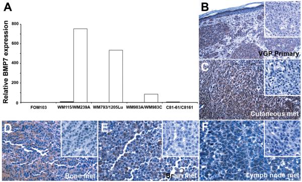
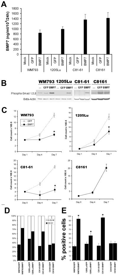
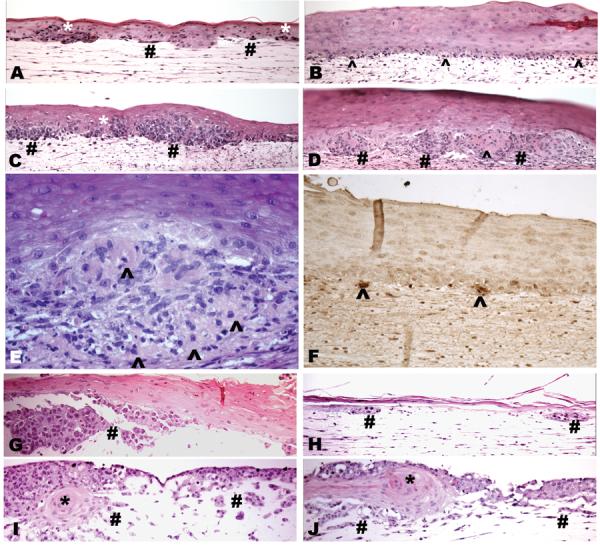
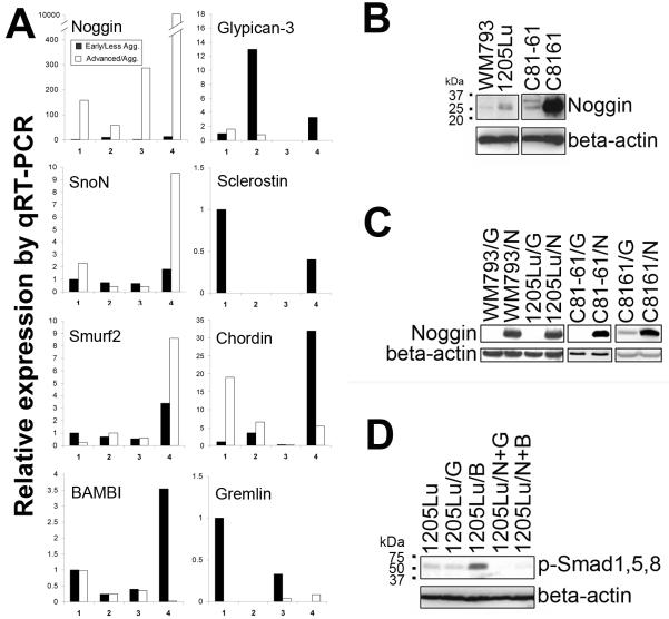
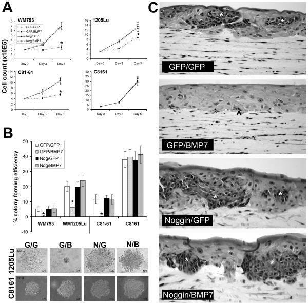
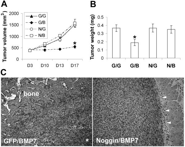
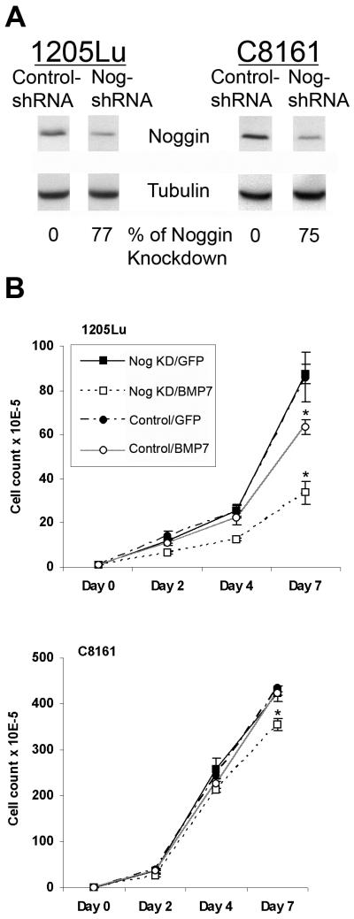
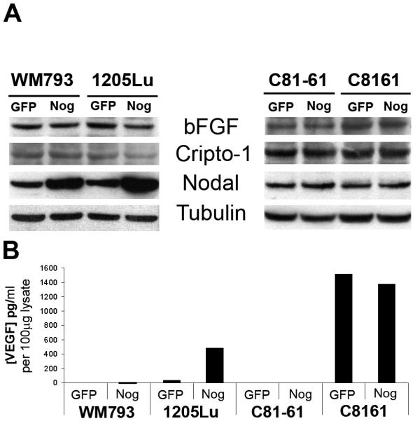
Bone morphogenetic proteins (BMPs) are members of the TGF-beta superfamily responsible for mediating a diverse array of cellular functions both during embryogenesis and in adult life. Previously, we reported that upregulation of BMP7 in human melanoma correlates with tumor progression. However, melanoma cells are either inhibited by or become resistant to BMP7 as a function of tumor progression, with normal melanocytes being most susceptible. Herein, real-time quantitative reverse transcriptase-polymerase chain reactions and western blotting revealed that the expression of BMP antagonist, Noggin, correlates with resistance to BMP7 in advanced melanoma cells. To test the hypothesis that coordinated upregulation of Noggin protects advanced melanoma cells from autocrine inhibition by BMP7, functional expression of Noggin in susceptible melanoma cells was achieved by adenoviral gene transfer. The Noggin-overexpressing cells exhibited a growth advantage in response to subsequent BMP7 transduction in vitro under anchorage-dependent and -independent conditions, in three-dimensional skin reconstructs, as well as in vivo in severe combined immunodeficient mice. In concordance, Noggin knockdown by lentiviral shRNA confers sensitivity to BMP7-induced growth inhibition in advanced melanoma cells. Our findings suggest that, like TGF-beta, BMP7 acts as an autocrine growth inhibitor in melanocytic cells, and that advanced melanoma cells may escape from BMP7-induced inhibition through concomitant aberrant expression of Noggin.


Figures

Figure 1
Figure 1
Expression of BMP7 correlates with melanoma progression in melanoma cell lines by qRT-PCR using normal melanocyte (FOM103) as control (A) and in situ by immunohistochemistry (B-F). Four isogenic cell pairs were analyzed by qRT-PCR, including: WM115 (primary melanoma, ■)/WM239A (metastatic melanoma, □); WM793 (primary melanoma, ■)/1205Lu (metastatic melanoma, □); WM983A (primary melanoma, ■)/WM983C (metastatic melanoma, □); and C81-61 (metastatic melanoma, ■)/C8161 (highly aggressive metastatic melanoma selected in experimental metastasis model in vivo, □). Paraffin sections of primary cutaneous melanoma (Magnification: 200X) and a panel of metastatic lesions in different organ sites (magnification: 400X), including skin (C), bone (D), brain (E), and lymph node (F), were subjected to immunohistochemistry as described in Materials and Methods. Comparing to the corresponding negative controls (inserts), BMP7 expression is evident in melanoma cells.
Figure 2
Figure 2
Biological consequences of functional expression of BMP7 in melanoma cells by adenoviral gene transfer. A. Secretion of transduced BMP7 by ELISA. Culture supernatants of transduced melanoma cells were subjected to ELISA using those of mock- and GFP-transduced cells as control. The levels of BMP7 expression were comparable among different cell lines averaging at ∼1000 ng/ml/106 cells/24 h. B. Transduced BMP7 activates R-Smad phosphorylation as determined by Western blotting. Using an Ab specific for phosphorylated Smads 1, 5, and 8, upregulation of phosphorylated R-Smads following BMP7 transduction in melanoma cells was observed with one exception (C8161 cells). The finding indicates that transduced BMP7 is biologically active and can function as an autocrine in melanoma by eliciting the Smad signaling cascade. C. Transduced BMP7 inhibits melanoma growth to different degrees in vitro in conventional culture. Early/less aggressive melanoma cells (WM793 and C81-61) were susceptible, demonstrating greater than 70% growth retardation in response to BMP7, while their highly aggressive variants (1205Lu and C8161) were relatively resistant with less than 50% inhibition. C8161, in particular, was completely resistant. Asterisks indicate statistically significant differences (p<0.05). D. Propidium iodide stain revealed that BMP7 transduction resulted in an increased percentage of cells resting at G0-G1 (solid column) comparing to that in Ad/GFP- and mock-infected cells. BMP7 drastically induced G0-G1 cell cycle arrest in susceptible melanoma cells (WM793 and C81-61), modestly in relatively resistant 1205Lu melanoma cells, and barely in resistant C8161 cells. E. BMP7 transduction induced early maker of apoptosis, phospho-H2B, in melanoma cells by flow cytometry. Comparing to GFP-transduced melanoma cells, a significant population of BMP7-transduced cells expressed phospho-H2B, particularly in WM793 and C81-61 cells. Asterisks indicate statistically significant differences (p<0.05).
Figure 3
Figure 3
Effects of transduced BMP7 on melanoma growth and invasion in 3D skin reconstructs. In susceptible primary melanoma cell line WM793, Ad/GFP-infected cells grow as large nests and single cells within the epidermis (*) and superficial dermis (#; A). Upon BMP7-transduction, WM793 cells show small clusters as well as single units at the dermal epidermal junction (B, ^) reactive to TUNNEL stain (F, ^). GFP tranfectants of the relative resistant isogenic cell line 1205Lu grow aggressively forming large tumor nests in the deep dermis (C, #), while the BMP7-transfectants (D, #) displayed extensive cell death (^) with morphological hallmarks of apoptosis (D and E, ^). Control GFP-transfected susceptible metastatic C81-61 melanoma cells grow aggressively as large nests and nodules within the dermis (G, #), whereas their BMP7-transduced counterparts only show small remnants of dermal tumor nests (H, #). In the resistant, highly aggressive C8161 melanoma cells, both Ad/GFP- (I) and Ad/BMP7-infected cells (J) grow aggressively into the dermis (#) and eventually also replace the epidermis with rare residual keratinocyte islands (I, J, *).
Figure 4
Figure 4
Noggin mediates resistance to BMP7. Noggin upregulation correlates with melanoma progression and resistance to BMP7 by qRT-PCR (A) and Western blotting (B). Isogenic cell pairs: 1. WM115 (primary melanoma, ■)/WM239A (metastatic melanoma, □); 2. WM793 (primary melanoma, ■)/1205Lu (metastatic melanoma, □); 3. WM983A (primary melanoma, ■)/WM983C (metastatic melanoma, □); 4. C81-61 (metastatic melanoma, ■)/C8161 (highly aggressive metastatic melanoma, □). C. Infection of susceptible melanoma cells with Noggin-expressing adenovirus and analysis by Western blotting (G: Ad/GFP-infected cells, N: Ad/Noggin-infected cells). D. Noggin expression blocks BMP7-induced phosphorylation of R-Smads. (G: Ad/GFP-infected cells, B: Ad/BMP7-infected cells, N+G: Ad/Noggin and Ad/GFP double infected cells, N+B: Ad/Noggin and Ad/BMP7 double infected cells.)
Figure 5
Figure 5
Noggin expression confers BMP7 resistance in susceptible melanoma cells in vitro. A. Conventional growth assay. Isogenic melanoma cell pairs (WM793/ 1205Lu, and C81-61/C8161) were sequentially infected with different combinations of adenoviral vectors and analyzed for growth rate in conventional culture conditions. BMP7 expression in control Ad/GFP-infected susceptible melanoma cell lines (WM793, and C81-61) results in marked growth inhibition, while in their Ad/Noggin-infected counterparts, cell growth is restored to the same rate as control Ad/GFP and Ad/GFP- double infected cells. Similar rescuing effect of Noggin is also observed in the relatively resistant 1205Lu cells. The resistant, highly aggressive melanoma cell line, C8161 remains resistant regardless of Noggin rescue. Asterisks indicate statistical significance (p<0.05). B. Colony formation in soft agar. Twenty-four h following double infection with different combinations of adenoviral vectors, melanoma cells were resuspended in 0.25% agar and seeded in triplicate wells at 6 × 10⁴ cells/well. Two weeks later, the colony-forming efficiency was determined as the percentage of cells forming colonies containing four or more cells. Ten random fields were examined for each condition. Data represent mean ± SD. Asterisks indicate significant difference (p<0.05). Representative micrographs of individual colonies of 1205Lu and C8161 are also shown. C. 3D skin reconstructs. Twenty-four h following double infection using various combinations of adenoviral vectors, susceptible WM793 primary melanoma cells were incorporated into 3D skin reconstructs. While Ad/GFP and Ad/BMP7-double infected cells result in scattered apoptotic cells at the dermal-epidermal junction and superficial dermis (^), the Ad/Noggin and Ad/BMP7-double infected cells grow as large tumor nests (*) within the epidermis and focally in the superficial dermis, a pattern similar to those of Ad/GFP and Ad/GFP-, and Ad/Noggin and Ad/GFP-infeected control.
Figure 6
Figure 6
Ectopic Noggin expression rescues tumorigenicity of Ad/BMP7-infected melanoma cells in vivo. Sixteen h after double infection, 1205Lu melanoma cells were injected subcutaneously in to SCID mice. Tumor sizes were monitored for 17 days (A) before the tumors were harvested, weighed (B), and processed for routine histology (C). Hematoxylin and Eosin stained slides show that the Ad/GFP and Ad//BMP7-double infected cells induce ectopic bone formation at the periphery of the tumor with central necrosis (*), while the Ad/Noggin and Ad/BMP7-double infected cells grow as large cutaneous nodules in delicate fibrous capsules (arrow heads) without evidence of heterotropic ossification.
Figure 7
Figure 7
Noggin knockdown in advanced melanoma cells confers sensitivity to BMP7-induced growth inhibition. A. Noggin knockdown by lentiviral shRNA. Stable cell lines expressing lentiviral shRNA against human Noggin and non-target control were subjected to Western blotting using tubulin as internal loading control. Above 75% Noggin knockdown efficiency is achieved as determined by densitometry in advanced 1205Lu and C8161 melanoma cells. B. Noggin knockdown renders advanced melanoma cells more sensitive to BMP7-induced growth inhibition. Upon infection by Ad/BMP7, 1205Lu and C8161 Noggin knockdown melanoma cells (□) show greater degrees of growth retardation compared to the their non-target control counterparts (○). Asterisks indicate statistically significant difference.
Figure 8
Figure 8
Effects of Noggin on melanoma growth promoting factors by Western blotting (A) and ELISA (B). Noggin transduction upregulates Nodal and VEGF in one isogenic melanoma cell pair WM793/1205Lu but not the other C81-61/C8161.

References

    1. Balemans W, Van Hul W. Extracellular regulation of BMP signaling in vertebrates: a cocktail of modulators. Dev Biol. 2002;250:231–250. - PubMed
    1. Whitman M. Smads and early developmental signaling by the TGFbeta superfamily. Genes Dev. 1998;12:2445–2462. - PubMed
    1. Botchkarev VA. Bone morphogenetic proteins and their antagonists in skin and hair follicle biology. J Invest Dermatol. 2003;120:36–47. - PubMed
    1. Hsu MY, Rovinsky S, Penmatcha S, et al. Bone morphogenetic proteins in melanoma: angel or devil? Cancer Metastasis Rev. 2005;24:251–263. - PubMed
    1. Howe JR, Sayed MG, Ahmed AF, et al. The prevalence of MADH4 and BMPR1A mutations in juvenile polyposis and absence of BMPR2, BMPR1B, and ACVR1 mutations. J Med Genet. 2004;41:484–491. - PMC - PubMed

Publication types

MeSH terms