. 2008 Aug;16(8):1450-8.
doi: 10.1038/mt.2008.127. Epub 2008 Jun 17.

Control of endothelial targeting and intracellular delivery of therapeutic enzymes by modulating the size and shape of ICAM-1-targeted carriers

Affiliations

Control of endothelial targeting and intracellular delivery of therapeutic enzymes by modulating the size and shape of ICAM-1-targeted carriers

Silvia Muro et al. Mol Ther. 2008 Aug.

Abstract

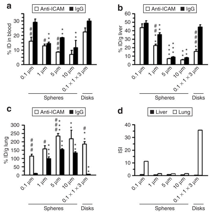
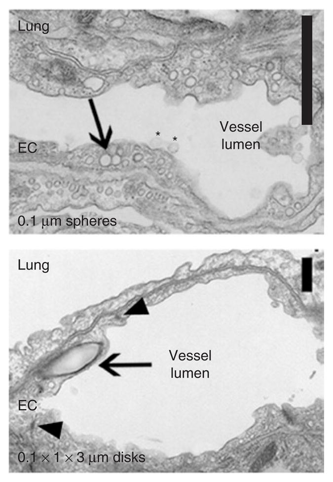
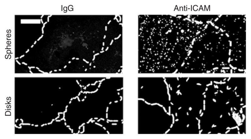
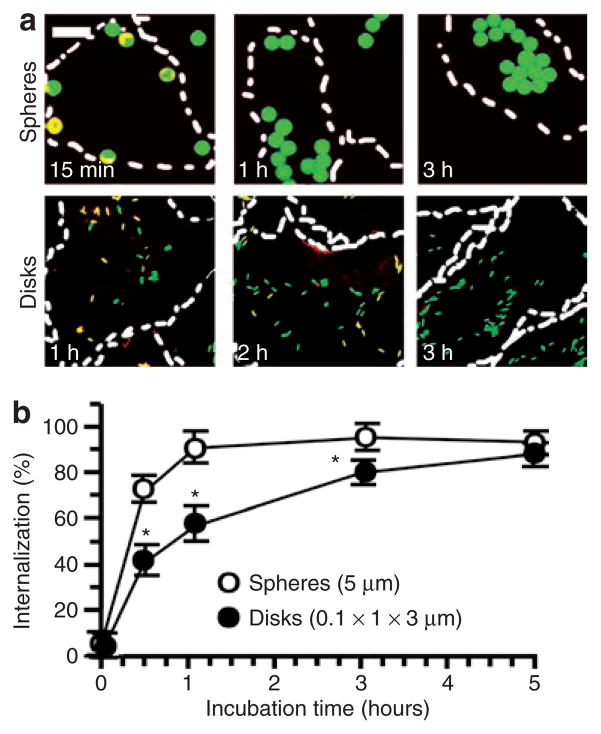
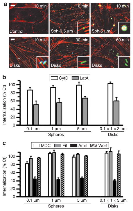
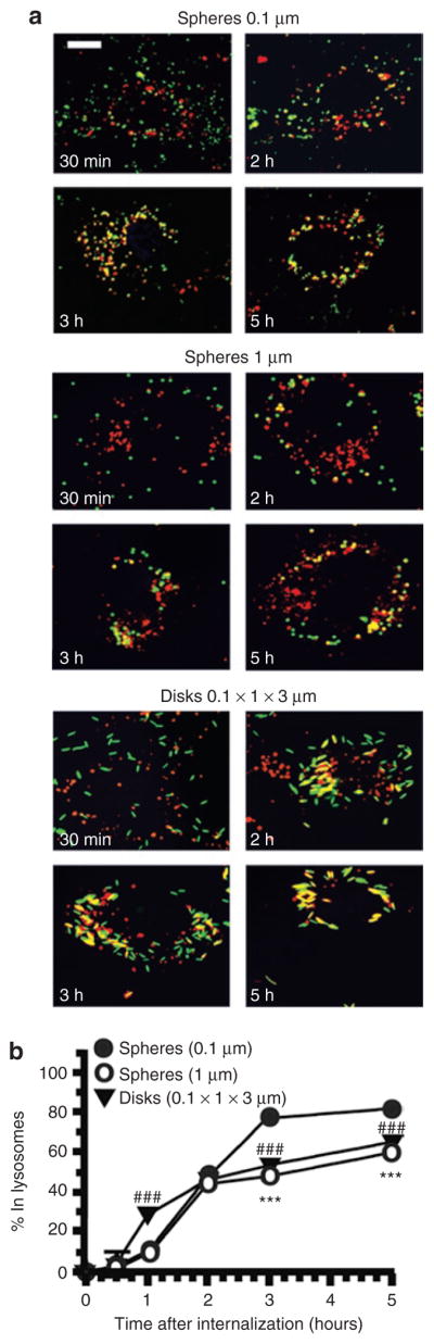
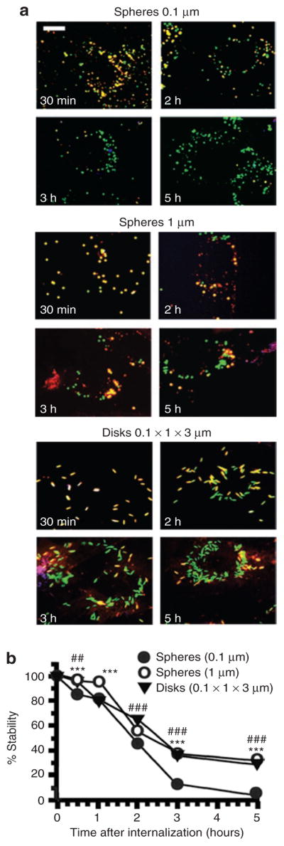
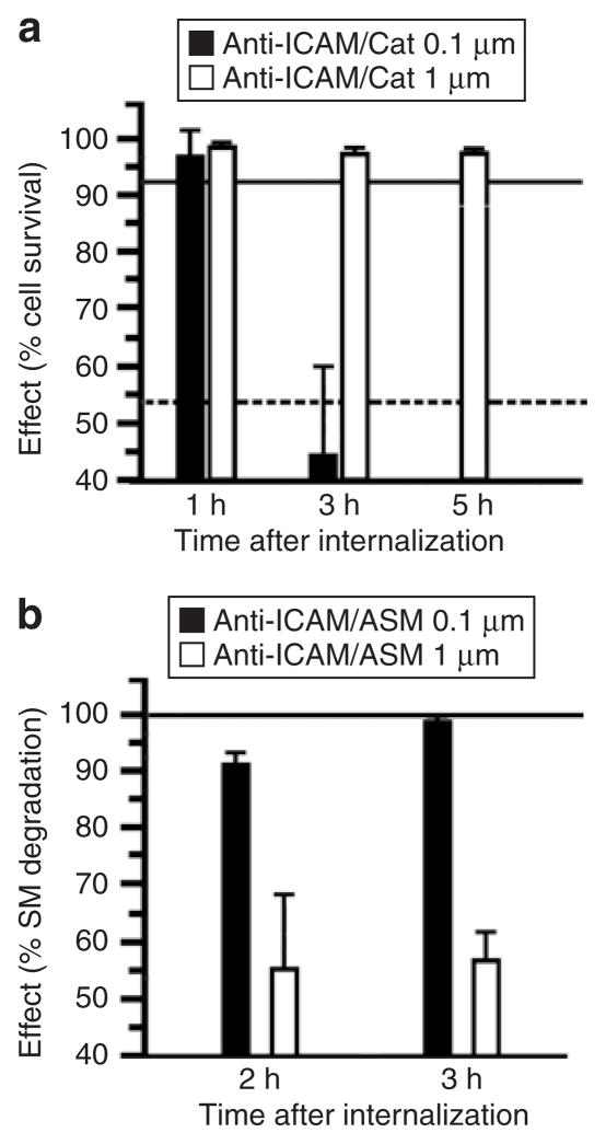
Endocytosis in endothelial cells (ECs) is important for many biomedical applications, including drug delivery by nano- and microscale carriers. However, little is known about how carrier geometry influences endothelial drug targeting, intracellular trafficking, and effects. We studied this using prototype polymer carriers of various sizes (0.1-10 mum) and shapes (spheres versus elliptical disks). Carriers were targeted to intercellular adhesion molecule 1 (ICAM-1), a transmembrane glycoprotein that is upregulated in many pathologies and used as a target for intraendothelial drug delivery. ECs internalized anti-ICAM-coated carriers of up to several microns in size via cell adhesion molecule-mediated endocytosis. This pathway is distinct from caveolar and clathrin endocytosis that operate for submicron-size objects. Carrier geometry was found to influence endothelial targeting in the vasculature, and the rate of endocytosis and lysosomal transport within ECs. Disks had longer half-lives in circulation and higher targeting specificity in mice, whereas spheres were endocytosed more rapidly. Micron-size carriers had prolonged residency in prelysosomal compartments, beneficial for endothelial antioxidant protection by delivered catalase. Submicron carriers trafficked to lysosomes more readily, optimizing effects of acid sphingomyelinase (ASM) enzyme replacement in a model of lysosomal storage disease. Therefore, rational design of carrier geometry will help optimize endothelium-targeted therapeutics.


Figures

Figure 1
Figure 1. Role of geometry in the pharmacokinetics and biodistribution of anti-ICAM carriers in mice
(a) Blood level of anti-ICAM (white bars) versus immunoglobulin G (IgG) (black bars) particles of various geometries (0.1, 1, 5, and 10 μm spheres, and 0.1 × 1 × 3 μm disks), calculated as percentage of injected dose (% ID) remaining in the circulation 1 minute after intravenous injection in C57BL/6 mice. (b) Liver uptake and (c) lung uptake (expressed as % ID per gram) of anti-ICAM (white bars) versus IgG (black bars) formulations, 30 minutes after injection. (d) The immunospecificity index (ISI) in liver (black bars) and lung (white bars) represents the anti-ICAM-to-IgG particle ratio, calculated as % ID/g in each of these tissues. Data are mean ± SEM (n ≥ 4 mice). *, Compares particles of any given micron-range size to 0.1 μm. #, compares anti-ICAM particles to IgG counterparts. * or #, P ≤ 0.05; ** or ##, P ≤ 0.01; *** or ###, P ≤ 0.001, by Student’s t-test. ICAM, intercellular adhesion molecule 1.
Figure 2
Figure 2. Internalization of anti-ICAM carriers by endothelial cells (ECs) in vivo
Transmission electron microscopy micrographs showing binding (asterisks) and internalization (arrows) of 0.1-μm anti-ICAM/spheres and 0.1 × 1 × 3 μm anti-ICAM/disks by pulmonary ECs 3 hours after injection. Intact cell junctions are marked by arrowheads. Scale bar = 1 μm. ICAM, intercellular-adhesion molecule 1.
Figure 3
Figure 3. Specific binding of anti-ICAM carriers to endothelial cells in culture
As an example, the fluorescent images show specific binding of fluorescein isothiocyanate–labeled 0.1-μm anti-ICAM spheres and 0.1 × 1 × 3 μm anti-ICAM disks versus immunoglobulin G (IgG) counterparts to tumor necrosis factor-α activated HUVECs (30 minutes incubation at 4 °C). Scale bar = 10 μm. The cells borders have been marked by a dashed line from phase-contrast images. ICAM, intercellular-adhesion molecule 1.
Figure 4
Figure 4. Role of geometry in the endocytosis of anti-ICAM carriers of by endothelial cells
(a) Fluorescence micrographs showing internalized fluorescein isothiocyanate–labeled (green) anti-ICAM/spheres (5 μm diameter) versus anti-ICAM/disks (0.1 × 1 × 3 μm) incubated with tumor necrosis factor-α activated HUVECs at 37 °C for the indicated time. Counterstaining with a Texas red secondary antibody reveals surface-accessible anti-ICAM particles (yellow). Dashed line = cell borders determined from phase-contrast images of cell monolayers. Scale bar = 10 μm. (b) Comparison of internalization kinetics of these anti-ICAM particle formulations, automatically quantified from fluorescence micrographs. Data are mean ± SEM (n ≥ 25 cells, two experiments). *Compares spheres to elliptical disks at any given time point. *, P ≤ 0.05, by Student’s t-test. ICAM, intercellular-adhesion molecule 1.
Figure 5
Figure 5. Mechanism of endocytosis of anti-ICAM carriers of various geometries
(a) Fluorescence microscopy showing formation of actin stress fibers (stained by red Alexa Fluor 594 phalloidin) upon incubation of activated HUVECs with fluorescein isothiocyanate–labeled anti-ICAM/spheres (0.5 and 5 μm diameter) or anti-ICAM/disks (0.1 × 1 × 3 μm) for the indicated time. Particles in the cell surface look blue due to counter-staining with blue Alexa Fluor 350 goat anti-mouse immunoglobulin G. Scale bar = 10 μm. (b) Internalization (1 hour) of anti-ICAM spherical particles of various sizes (0.1, 1, and 5 μm diameter) and elliptical disks (0.1 × 1 × 3 μm) in the presence of two pharmacological inhibitors of actin filaments (0.5 μmol/l cytochalasin D or CytD, and 0.1 μmol/l latrunculin A or LatA). (c) The effects of pharmacological inhibitors of clathrin-coated pits (50 μmol/l monodansyl cadaverine, MDC), caveolar-mediated endocytosis (1 μg/ml filipin, Fil), a common inhibitor to macropinocytosis and CAM endocytosis (3 mmol/l amiloride, Amil), and a macropinocytosis inhibitor (0.5 μmol/l Wortmannin, Wort), were tested as in (b). Data are mean ± SEM (n ≥ 25 cells, two experiments). Calculated with respect to control cells (%Ct). ICAM, intercellular adhesion molecule 1.
Figure 6
Figure 6. Role of geometry on the intracellular trafficking of anti-ICAM carriers
(a) Fluorescence micrographs showing trafficking of fluorescein isothiocyanate–labeled (green) anti-ICAM carriers of various geometries (0.1 and 1 μm spheres, and 0.1 × 1 × 3 μm disks) to Texas red dextran prelabeled lysosomes (red). Lysosomal colocalization is visualized as yellow. Scale bar = 10 μm. (b) Trafficking of anti-ICAM carriers to lysosomes was calculated as percent colocalization of these fluorescent markers determined by microscopy at the indicated time. Data are mean ± SEM (n > 25 cells, two experiments). Low-value error bars are masked by symbols in the graph. *, Compares 1 μm spheres to 0.1 μm particles at any given time point. #, compares 0.1 × 1 × 3 μm disks to 0.1 μm particles at any given time point. * or #, P ≤ 0.05; ** or ##, P ≤ 0.01; *** or ###, P ≤ 0.001, by Student’s t-test. ICAM, intercellular-adhesion molecule 1.
Figure 7
Figure 7. Role of geometry on the intracellular stability of anti-ICAM carriers
(a) Fluorescence micrographs showing fluorescein isothiocyanate–labeled (green) anti-ICAM carriers of various geometries (0.1 and 1 μm spheres, and 0.1 × 1 × 3 μm disks), after counterstaining surface-bound carriers with a blue Alexa Fluor 350 secondary antibody to anti-ICAM. The stability of anti-ICAM protein counterpart in the internalized carrier (green) was then assessed by anti-ICAM immunodetection using a Texas red–conjugated secondary antibody after cell permeabilization. Hence, yellow denotes stability of anti-ICAM carrier counterpart. Scale bar = 10 μm. (b) Proteolytic degradation of anti-ICAM protein counterpart onto the particles quantified from fluorescence micrographs. Data are mean ± SEM (n > 25 cells, two experiments). Low-value error bars are masked by symbols in the graph. *, compares 1 μm spheres to 0.1 μm particles at any given time point. #, Compares 0.1 × 1 × 3 μm disks to 0.1 μm particles at any given time point. * or #, P ≤ 0.05; ** or ##, P ≤ 0.01; *** or ###, P ≤ 0.001, by Student’s t-test. ICAM, intercellular-adhesion molecule 1.
Figure 8
Figure 8. Role of geometry on the functional therapeutic activity of anti-ICAM carriers
(a) Induction of oxidative injury in HUVECs with 5 mmol/l H2O2 after delivery of antioxidant catalase by 0.1 versus 1 μm spherical anti-ICAM particles. Cell survival was estimated by labeling HUVECs with Live/Dead assay and fluorescent imaging. Data are mean ± SEM (n ≥ 500 cells/condition). The continuous and dashed lines in the graph represent survival levels of noninjured cells and H2O2-treated cells, respectively, tested after incubation with control 0.1 μm anti-ICAM particles. (b) Aberrant storage of sphingomyelin (SM), typical of the lysosomal storage disorder type A and B Niemann–Pick disease, was induced in HUVECs by treatment with 50 μmol/l imipramine. SM was labeled in these deficient cells by incubation for 16 hours at 37 °C with a BODIPY-FLC12-SM analog. Reduction of SM within these intracellular compartments was imaged after internalization of recombinant acid sphingomyelinase, delivered by either 0.1 versus 1 μm spherical anti-ICAM carriers. Intracellular level of SM was quantified by fluorescence microscopy, and normalized to SM levels in normal HUVECs versus diseased HUVECs before enzyme replacement. Data are mean ± SEM (n ≥ 10 cells, two assays). ICAM, intercellular-adhesion molecule 1.

References

    1. Song BW, Vinters HV, Wu D, Pardridge WM. Enhanced neuroprotective effects of basic fibroblast growth factor in regional brain ischemia after conjugation to a blood-brain barrier delivery vector. J Pharmacol Exp Ther. 2002;301:605–610. - PubMed
    1. Muzykantov V. Targeting drugs to pulmonary endothelium. Expert Opinion Drug Delivery. 2005;2:909–926. - PubMed
    1. Oh P, Li Y, Yu J, Durr E, Krasinska KM, Carver LA, et al. Subtractive proteomic mapping of the endothelial surface in lung and solid tumours for tissue-specific therapy. Nature. 2004;429:629–635. - PubMed
    1. Wilson A, He F, Li J, Ma Z, Pitt B, Li S. Targeted delivery of therapeutic oligonucleotides to pulmonary circulation. Adv Genet. 2005;54:21–41. - PubMed
    1. Molema G. Tumor vasculature directed drug targeting: applying new technologies and knowledge to the development of clinically relevant therapies. Pharm Res. 2002;19:1251–1258. - PubMed

Publication types

MeSH terms