. 2008 Jun 18;3(6):e2518.
doi: 10.1371/journal.pone.0002518.

Suppression of tumor growth and angiogenesis by a specific antagonist of the cell-surface expressed nucleolin

Affiliations

Suppression of tumor growth and angiogenesis by a specific antagonist of the cell-surface expressed nucleolin

Damien Destouches et al. PLoS One. .

Abstract

Background: Emerging evidences suggest that nucleolin expressed on the cell surface is implicated in growth of tumor cells and angiogenesis. Nucleolin is one of the major proteins of the nucleolus, but it is also expressed on the cell surface where is serves as a binding protein for variety of ligands implicated in cell proliferation, differentiation, adhesion, mitogenesis and angiogenesis.

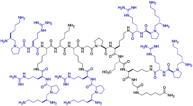
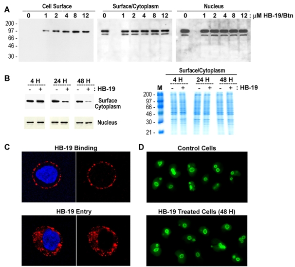
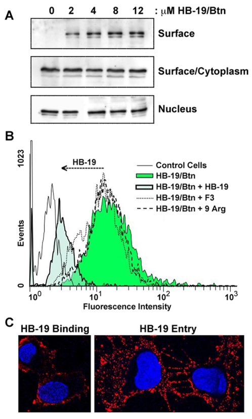
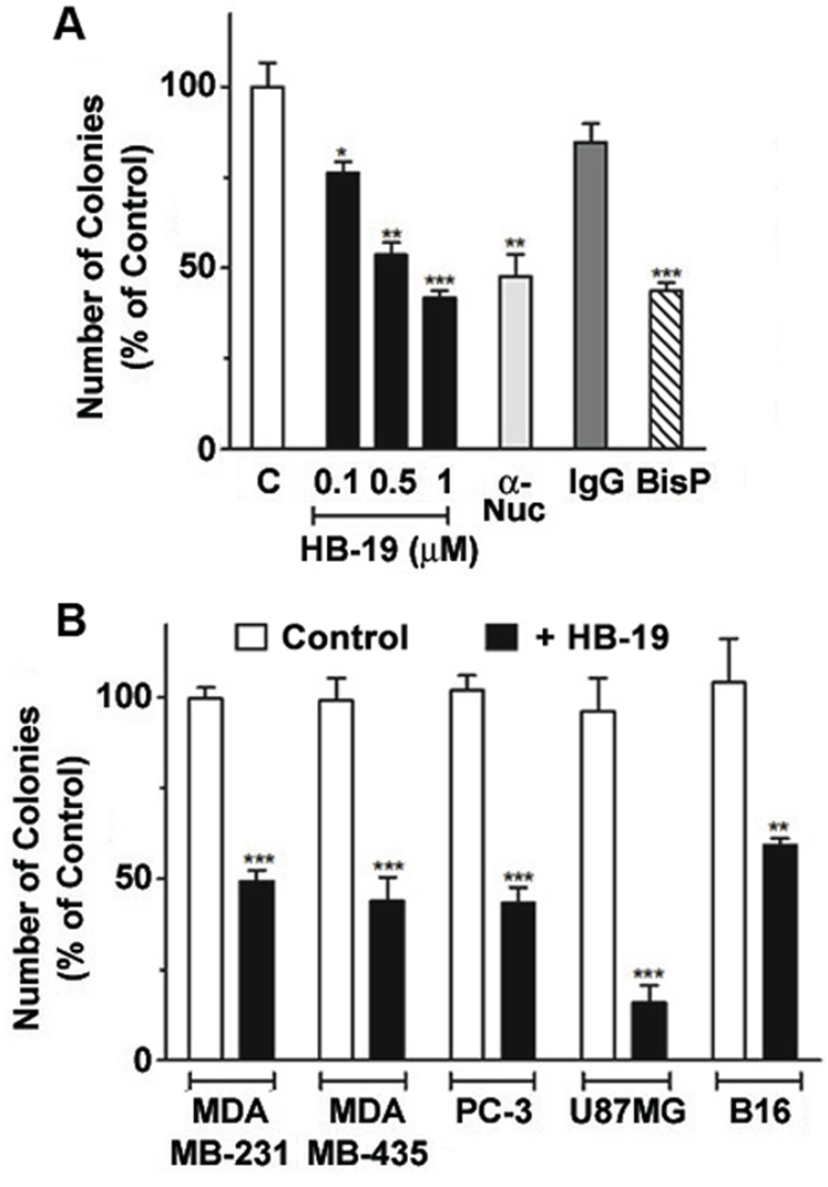
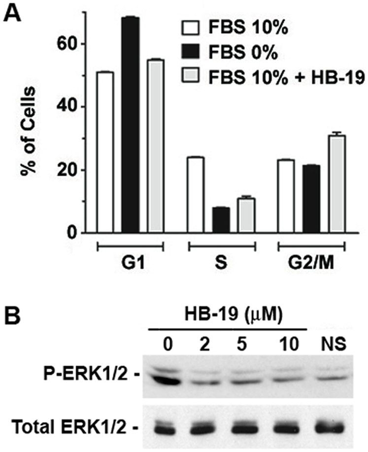
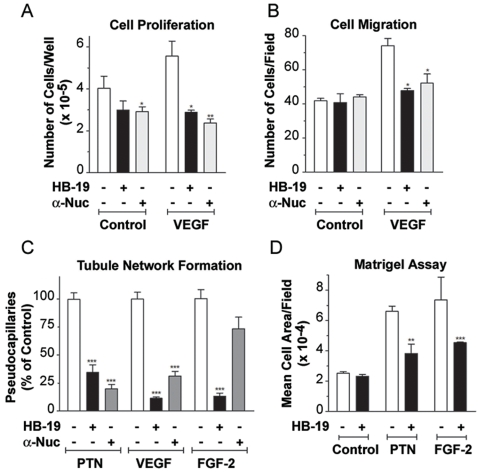
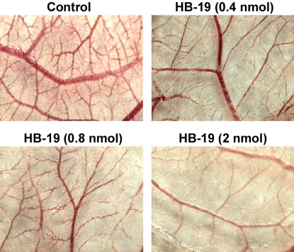
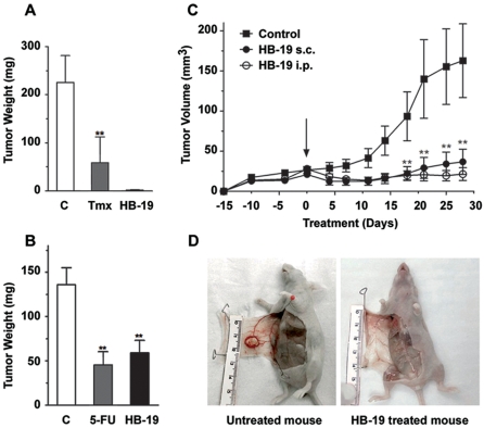
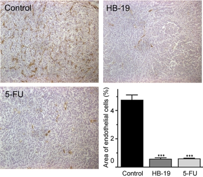
Methodology/principal findings: By using a specific antagonist that binds the C-terminal tail of nucleolin, the HB-19 pseudopeptide, here we show that the growth of tumor cells and angiogenesis are suppressed in various in vitro and in vivo experimental models. HB-19 inhibited colony formation in soft agar of tumor cell lines, impaired migration of endothelial cells and formation of capillary-like structures in collagen gel, and reduced blood vessel branching in the chick embryo chorioallantoic membrane. In athymic nude mice, HB-19 treatment markedly suppressed the progression of established human breast tumor cell xenografts in nude mice, and in some cases eliminated measurable tumors while displaying no toxicity to normal tissue. This potent antitumoral effect is attributed to the direct inhibitory action of HB-19 on both tumor and endothelial cells by blocking and down regulating surface nucleolin, but without any apparent effect on nucleolar nucleolin.

Conclusion/significance: Our results illustrate the dual inhibitory action of HB-19 on the tumor development and the neovascularization process, thus validating the cell-surface expressed nucleolin as a strategic target for an effective cancer drug. Consequently, the HB-19 pseudopeptide provides a unique candidate to consider for innovative cancer therapy.


Conflict of interest statement

Competing Interests: The authors have declared that no competing interests exist.

Figures

Figure 1
Figure 1. Molecular structure of the multivalent HB-19 pseudopeptide.
HB-19 presents pentavalently the pseudo-tripeptide Lysψ(CH2N)-Pro-Arg coupled to a template (H2NLys-Lys-Lys-Gly-Pro-Lys-Glu-Lys-AhxCONH2); ψ(CH2N) stands for a reduced peptide bond between Lys and Pro residues. The tripeptide is assembled on the α-NH2 of the template and the ε-NH2 groups of the four lysine residues indicated in bold. No apparent modification is observed for the HPLC profile of HB-19 after five days of incubation at 37°C in normal human serum, thus illustrating its resistance to degradation by serum proteases.
Figure 2
Figure 2. HB-19 binds nucleolin expressed on the surface of tumor cells and causes reduction of cytoplasmic/surface but not nuclear nucleolin.
(A) HB-19/Btn forms a stable complex with nucleolin expressed on the surface of cells. MDA-MB-231 cells were incubated (45 min, 20°C) with 0, 1, 2, 4, 8 and 12 µM of HB-19/Btn before preparation of surface/cytoplasmic (i.e., nucleus-free) and nuclear extracts , . Samples of surface nucleolin (purified using surface/cytoplasmic extracts from 2×10⁷ cells) and crude surface/cytoplasmic and nuclear extracts (from 2×10⁶ cells) were analyzed by immunoblotting for the detection of nucleolin using mAb D3. (B) Specific reduction of surface/cytoplasmic nucleolin in HB-19 treated cells. MDA-MB-231 cells were cultured (at 37°C) with 10 µM of HB-19 for 4, 24 and 48 hours before preparation of cytoplasmic and nuclear extracts. Material from 2×10⁶ cells (Untreated or HB-19 treated cells as indicated − or + signs, respectively) was analyzed by immunoblotting for the detection of nucleolin. Sections of the gel at the position of the nucleolin bands are presented. The right panel shows the profile of proteins in the Nuclear-free cell extract (Surface/Cytoplasm) of the PAGE-SDS gel stained with Brilliant Blue G-Colloidal Concentrate. Lane M shows the electrophoretic mobility of protein markers. The intensity of nucleolin protein bands quantified by using the NIH image software indicated 75% and 93% reduction of surface/cytoplasmic nucleolin in HB-19 treated cells at 24 and 48 hours, respectively, compared to the corresponding untreated cells. (C) HB-19 binds the cell surface and enters in the cytoplasm but not the nucleus. MDA-MB-231 cells were incubated at 20°C (for HB-19 Binding) or 37°C (for HB-19 Entry) with 5 µM of HB-19/Btn for 1 hour before fixation in PFA or PFA-Triton, respectively. Fixed cells were successively incubated with rabbit anti-biotin and goat Texas Red-conjugated anti-rabbit IgG and the nuclei were colored with DAPI. Scans corresponding to the cross-section of cells are shown, each with or without the DAPI. (D) Nucleolar nucleolin is not affected in HB-19 treated cells. MDA-MB-231 cells cultured in the absence (Control Cells) or presence of 10 µM of HB-19 for 48 hours (HB-19 Treated Cells) were fixed in PFA-Triton and processed for fluorescence microscopy. Nucleolin was revealed by mAb D3 and FITC-conjugated goat anti-mouse IgG.
Figure 3
Figure 3. Specific binding and entry of HB-19 in endothelial cells.
(A) HB-19/Btn forms a stable complex with nucleolin expressed on the surface of HUVECs. Cells were incubated (45 min, 20°C) with 0, 1, 2, 4, 8 and 12 µM of HB-19/Btn before preparation of surface/cytoplasmic and nuclear extracts and recovery of surface nucleolin from the surface/cytoplasmic extracts as in Figure 2A. Material extracted from 2×10⁷ and 2×10⁶ cells was analyzed in panels Surface and Surface/Cytoplasm or Nucleus, respectively. Immunoblotting was with the anti-nucleolin mAb D3. (B) The specific binding of HB-19/Btn to HUVECs. The binding of 1 µM HB-19/Btn was studied at 4 °C by FACScan analysis. To show the specificity of binding, the reaction was carried out in the absence or presence of 50 µM HB-19, F3, or 9Arg peptides. The ordinate gives the relative cell number, whereas the abscissa gives the relative fluorescence intensity. (C) HB-19 entry in HUVECs. Cells were incubated with 5 µM of HB-19/Btn (1 hour, 37°C) before fixation in PFA (for surface binding) or PFA-Triton (for entry), respectively. Cells were then successively incubated with rabbit anti-biotin antibodies and goat Texas Red-conjugated anti-rabbit IgG, and processed for confocal microscopy. The scans of cells toward the middle cell layer are presented (with the DAPI stained nuclei).
Figure 4
Figure 4. HB-19 inhibits colony formation in soft agar by tumor cell lines.
(A) MDA-MB-231 cells in culture medium were seeded in triplicate in the absence (histogram C) or presence of 0.1, 0.5 and 1 of µM HB-19, or 0.1 µM of the anti-nucleolin mAb MS-3 or control IgG, or 10 µM bisphosphonate (BisP). (B) Various tumor cell lines (as indicated) in culture medium were seeded in triplicate in the absence (histogram Control) or presence of 5 µM HB-19. After 10–21 days, colonies with diameters greater than 50 µm were scored as positive. Statistical significance: *0.01
Figure 5
Figure 5. Cell cycle perturbations induced by HB-19 treatment.
(A) Analysis of cell cycle parameters in HB-19 treated cells. MDA-MB-231 cells were cultured for 48 hours in medium without FBS (starvation) or in medium containing 10% FBS supplemented or not with 10 µM HB-19. DNA synthesis was quantified after BrdU incorporation and staining with anti-BrdU antibody and 7-AAD, by FACScan analysis. The histograms indicate the relative amount of cells in G1, S and G2/M cell phases. (B) HB-19 treatment inhibits serum-induced phosphorylation of ERK1/2. Serum starved MDA-MB-231 cells were stimulated with 10% FBS in the absence or presence of 2, 5, and 10 µM of HB-19. Five minutes after serum stimulation, cells were lysed directly in electrophoresis sample buffer and processed for immunoblotting using anti-phospho-p42/44 ERK1/2 and anti-p42/44 ERK antibodies. NS stands for non-stimulated cells.
Figure 6
Figure 6. Inhibition of in vitro and in vivo angiogenesis by HB-19.
(A) HB-19 inhibits proliferation of HUVECs. Twenty-four hours after seeding HUVECs in 2% FBS, cells were stimulated by 0.25 nM VEGF165, in the absence or presence of 1 µM HB-19 or 0.1 µM anti-nucleolin mAb MS-3 as indicated. After 72 h, cell number was determined by crystal violet staining . The data are reported as the mean of triplicate samples. (B) HB-19 inhibits migration of HUVECs. Cell migration was studied using a modified Boyden chamber. Cells were incubated (4 h at 37°C) with VEGF165, HB-19, or anti-nucleolin mAb MS-3 as above. Cells that migrated through the pore to the lower filter surface were counted and are shown as number per microscopic field at ×100 magnification. The data are the mean of three independent high-power fields/well performed in duplicate in two independent experiments. (C) HB-19 inhibits tubular network structure formation in collagen. Aortic endothelial (ABAE) cells in the absence or presence of 3 nM PTN, 0.5 nM VEGF165, and 3 nM FGF-2 were seeded on a three-dimensional collagen gel in complete medium. Treatment with HB-19 or anti-nucleolin mAb MS-3 was for 3 days after cell plating. Tubular network structures were quantified using phase contrast microscopy (×100). The ordinate gives the number of pseudocapillaries corresponding to the means of three randomly chosen fields/well from three wells. (D) HB-19 inhibits ex vivo angiogenic activity of growth factors in a Matrigel plug model. Liquid Matrigel was subcutaneously injected into the flank of Swiss mice in the absence or presence of 5 nM PTN, 10 nM FGF-2, and 1 µM HB-19. Quantification of endothelial cell invasion into the Matrigel was determined and is expressed as a mean of five fields per section from 3 Matrigel-plug sections per mouse. The results are expressed as the mean of five mice per group. Statistical significance: *p<0.05, **p<0.01, ***p<0.001.
Figure 7
Figure 7. Inhibition of in vivo angiogenesis by HB-19 in chick embryo chorioallantoic membrane (CAM) assay.
Macroscopic observation of the angiogenic response induced by gelatin sponges (1 cm2), soaked with 40 µl of PBS (Control) or 40 µl PBS containing HB-19 at concentrations of 10, 20 and 50 µM (corresponding to 0.4, 0.8 and 2 nmols of HB-19, respectively). After 48 h, CAMs were fixed and excised from the eggs . Photographs were taken and the total length of the vessels was measured using the Image PC image-analysis software (Scion Corporation, USA). Each sample was tested three times using 15–20 eggs for each data point. The relative percent inhibition (P<0.001) of angiogenesis compared to the control was 27, 36 and 51% in the presence of 0.4, 0.8 and 2 nmols of HB-19, respectively.
Figure 8
Figure 8. HB-19 inhibits tumor growth in the nude mice.
(A) HB-19 inhibits the growth of MDA-MB-231 tumor-cell xenografts. Cells (2×10⁶) were injected subcutaneously into the right flank of female nude mice. Two weeks later, mice with a palpable tumor of approximately 40 mm3 in volume were randomly separated into three groups (n = 5) and were given peritumoral injections 3 times/week of 0.1 ml PBS alone (Control), HB-19 (5 mg/kg), or Tamoxifen (Tmx) 10 mg/kg) for 6 weeks. (B) HB-19 inhibits the growth of MDA-MB-435 tumor-cell xenografts. Cells (1×10⁶) were injected in the mammary fat pad of female nude mice. Two weeks later, mice with a palpable tumor were randomly separated into three groups (n = 10) and were given intraperitoneal injections 3 times/week of 0.1 ml PBS alone (Control), HB-19 (5 mg/kg), or 5-fluouracil (5-FU, 40 mg/kg) for 8 weeks. At the end of each experiment (in A and B), mice were sacrificed and the tumors were excised and weighed. The results are presented as the mean weight ±standard deviation (±S.D.) obtained from the number of mice in each group. (C, D) Inhibition of tumor development in mice treated by intraperitoneal (i.p.) and subcutaneous (s.c.) administration of HB-19. MDA-MB-231 tumor bearing mice in three groups (n = 10) were treated with HB-19 (10 mg/kg) by i.p. or s.c. injections, 3 times/week for 28 days. The arrow at day 0 shows initiation of HB-19 treatment. Panel D shows MDA-MB-231 tumor bearing mice, untreated control and HB-19 treated (i.p. injection). Statistical significance: *p<0.05, **p<0.01, ***p<0.001.
Figure 9
Figure 9. Reduced density of blood vessels in HB-19 treated tumor-bearing mice.
Sections of tumors recovered from mice, untreated control and treated with either HB-19 or 5-FU (experiment described in Figure 8B) were stained with antibodies against the CD31 endothelial marker and analyzed by fluorescence microscopy. Representative macroscopic image (magnification 200×) from each group of mice shows the marked reduction of blood vessels in the tumors recovered from HB-19 or 5-FU treated animals. Angiogenesis was quantified by image analysis of CD31-labeled endothelial cells. The graph shows the mean areas ±standard deviation obtained from control and treated mice. ***p<0.001.

Similar articles

Cited by

References

    1. Srivastava M, Pollard HB. Molecular dissection of nucleolin's role in growth and cell proliferation: new insights. FASEB J. 1999;13:1911–1922. - PubMed
    1. Storck S, Shukla M, Dimitrov S, Bouvet P. Functions of the histone chaperone nucleolin in diseases. Subcell Biochem. 2007;41 - PubMed
    1. Hovanessian AG, Puvion-Dutilleul F, Nisole S, Svab J, Perret E, et al. The cell-surface-expressed nucleolin is associated with the actin cytoskeleton. Exp Cell Res. 2000;261:312–328. - PubMed
    1. Carpentier M, Morelle W, Coddeville B, Pons A, Masson M, et al. Nucleolin undergoes partial N- and O-glycosylations in the extranuclear cell compartment. Biochemisty. 2005;44:5804–5814. - PubMed
    1. Semenkovich CF, Ostlund REJ, Olson MO, Yang JW. A protein partially expressed on the surface of HepG2 cells that binds lipoproteins specifically is nucleolin. Biochemistry. 1990;29:9708–9713. - PubMed

Publication types

MeSH terms