

Transferrin fails to provide protection against Fas-induced hepatic injury in mice with deletion of functional transferrin-receptor type 2
- PMID: 18561026
- PMCID: PMC2574612
- DOI: 10.1007/s10495-008-0233-6
Transferrin fails to provide protection against Fas-induced hepatic injury in mice with deletion of functional transferrin-receptor type 2
Abstract
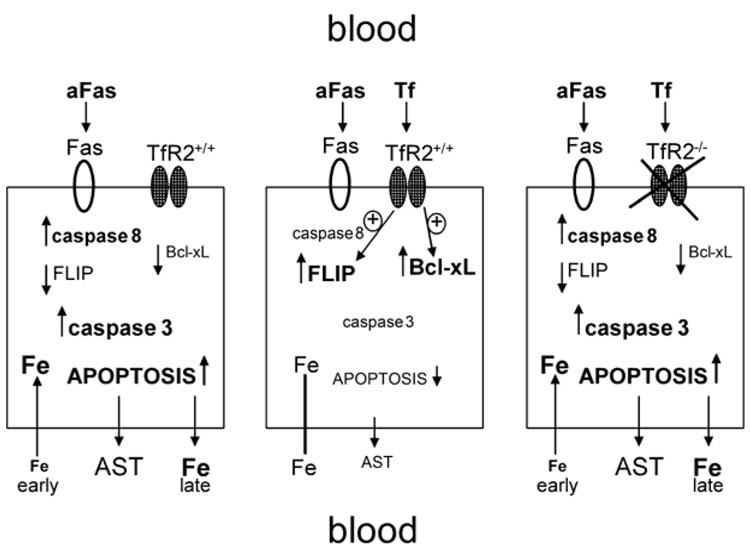
We reported previously that Fas-induced hepatic failure in normal mice was attenuated or prevented by exogenous transferrin (Tf), particularly apoTf. Here we show in C57BL6J/129 mice with genetic inactivation of transferrin receptor 2 (TfR2(Y245X)), that Fas-induced hepatotoxicity (apoptosis; rise in plasma aspartate aminotransferase (AST) levels) was comparable to that in wild-type mice, but was not modified by pretreatment with Tf. Rises in plasma AST were preceded by a decline in serum iron levels. AST elevations and iron declines were more profound in female than in male mice. Female mice also showed higher baseline levels of Bcl-xL in hepatocytes, which declined significantly upon treatment with agonistic anti-Fas antibody. These data confirm the cytoprotective function of Tf, and show a novel property of TfR2. Both apoptotic Fas responses and cytoprotective effects of Tf were associated with significant shifts in plasma iron levels, which quantitatively differed between male and female mice.
Conflict of interest statement
Figures


Similar articles
-
Protection of human and murine hepatocytes against Fas-induced death by transferrin and iron.Apoptosis. 2006 Jan;11(1):79-87. doi: 10.1007/s10495-005-3086-2. Apoptosis. 2006. PMID: 16374550
-
Prevention of Fas-mediated hepatic failure by transferrin.Lab Invest. 2004 Mar;84(3):342-52. doi: 10.1038/labinvest.3700035. Lab Invest. 2004. PMID: 14704719
-
Disruption of hemochromatosis protein and transferrin receptor 2 causes iron-induced liver injury in mice.Hepatology. 2012 Aug;56(2):585-93. doi: 10.1002/hep.25689. Epub 2012 Jun 11. Hepatology. 2012. PMID: 22383097
-
Transferrin and transferrin receptors update.Free Radic Biol Med. 2019 Mar;133:46-54. doi: 10.1016/j.freeradbiomed.2018.06.037. Epub 2018 Jun 30. Free Radic Biol Med. 2019. PMID: 29969719 Review.
-
Neuroimmunomodulation and aging: a role for transferrin and the hypothalamus/thymus axis.Curr Aging Sci. 2013 Feb;6(1):21-8. doi: 10.2174/1874609811306010004. Curr Aging Sci. 2013. PMID: 23895519 Review.
Cited by
-
Iron overload, hematopoietic cell transplantation, and graft-versus-host disease.Leuk Lymphoma. 2009 Oct;50(10):1566-72. doi: 10.1080/10428190903144659. Leuk Lymphoma. 2009. PMID: 19863335 Free PMC article. Review.
-
Allogeneic transplantation, Fas signaling, and dysregulation of hepcidin.Biol Blood Marrow Transplant. 2013 Aug;19(8):1210-9. doi: 10.1016/j.bbmt.2013.05.012. Epub 2013 May 22. Biol Blood Marrow Transplant. 2013. PMID: 23707854 Free PMC article.
-
Transplantation of allogeneic T cells alters iron homeostasis in NOD/SCID mice.Blood. 2009 Feb 19;113(8):1841-4. doi: 10.1182/blood-2008-09-178517. Epub 2008 Dec 23. Blood. 2009. PMID: 19109230 Free PMC article.
-
Viral infection, inflammation and schizophrenia.Prog Neuropsychopharmacol Biol Psychiatry. 2013 Apr 5;42:35-48. doi: 10.1016/j.pnpbp.2012.02.001. Epub 2012 Feb 10. Prog Neuropsychopharmacol Biol Psychiatry. 2013. PMID: 22349576 Free PMC article. Review.
-
The intracellular trafficking pathway of transferrin.Biochim Biophys Acta. 2012 Mar;1820(3):264-81. doi: 10.1016/j.bbagen.2011.09.009. Epub 2011 Sep 22. Biochim Biophys Acta. 2012. PMID: 21968002 Free PMC article. Review.
References
-
- Pierpaoli W, Lesnikov V, Lesnikova M, Arrighi S. Donor-derived plasma transferrin facilitates the engraftment of xenogeneic (rat) bone marrow in irradiated mice. Bone Marrow Transplant. 1996;18:203–207. [published erratum appears in Bone Marrow Transplant 1996 Dec;18(6):1201] - PubMed
-
- Weinzimer SA, Gibson TB, Collett-Solberg PF, Khare A, Liu B, Cohen P. Transferrin is an insulin-like growth factor-binding protein-3 binding protein. J Clin Endocrinol Metab. 2001;86:1806–1813. - PubMed
-
- Fassl S, Leisser C, Huettenbrenner S, Maier S, Rosenberger G, Strasser S, Grusch M, Fuhrmann G, Leuhuber K, Polgar D, Stani J, Tichy B, Nowotny C, Krupitza G. Transferrin ensures survival of ovarian carcinoma cells when apoptosis is induced by TNFalpha, FasL, TRAIL, or Myc. Oncogene. 2003;22:8343–8355. - PubMed
-
- Lesnikov V, Lesnikova M, Shulman HM, Arrighi S, Pierpaoli W, Deeg HJ. In vivo cytoprotective and immunomodulatory effects of transferrin. Research Advances in Blood. 2001;1:169–178.
-
- Aracena P, Aguirre P, Munoz P, Nunez MT. Iron and glutathione at the crossroad of redox metabolism in neurons. Biological Research. 2006;39:157–165. - PubMed
Publication types
MeSH terms
Substances
Grants and funding
- R01 CA074131/CA/NCI NIH HHS/United States
- R01 HL082941/HL/NHLBI NIH HHS/United States
- HL082941/HL/NHLBI NIH HHS/United States
- DK056465/DK/NIDDK NIH HHS/United States
- P30 DK056465/DK/NIDDK NIH HHS/United States
- P01 HL036444/HL/NHLBI NIH HHS/United States
- R01 CA023226/CA/NCI NIH HHS/United States
- HL036444/HL/NHLBI NIH HHS/United States
- R01 DK063016/DK/NIDDK NIH HHS/United States
- CA074131/CA/NCI NIH HHS/United States
- R37 CA023226/CA/NCI NIH HHS/United States
- DK063016/DK/NIDDK NIH HHS/United States
- CA023226/CA/NCI NIH HHS/United States
LinkOut - more resources
Full Text Sources
Research Materials
Miscellaneous
