. 2008 Sep 15;310(6):520-33.
doi: 10.1002/jez.b.21224.

Effects of nerve injury and segmental regeneration on the cellular correlates of neural morphallaxis

Affiliations

Effects of nerve injury and segmental regeneration on the cellular correlates of neural morphallaxis

Veronica G Martinez et al. J Exp Zool B Mol Dev Evol. .

Abstract

Functional recovery of neural networks after injury requires a series of signaling events similar to the embryonic processes that governed initial network construction. Neural morphallaxis, a form of nervous system regeneration, involves reorganization of adult neural connectivity patterns. Neural morphallaxis in the worm, Lumbriculus variegatus, occurs during asexual reproduction and segmental regeneration, as body fragments acquire new positional identities along the anterior-posterior axis. Ectopic head (EH) formation, induced by ventral nerve cord lesion, generated morphallactic plasticity including the reorganization of interneuronal sensory fields and the induction of a molecular marker of neural morphallaxis. Morphallactic changes occurred only in segments posterior to an EH. Neither EH formation, nor neural morphallaxis was observed after dorsal body lesions, indicating a role for nerve cord injury in morphallaxis induction. Furthermore, a hierarchical system of neurobehavioral control was observed, where anterior heads were dominant and an EH controlled body movements only in the absence of the anterior head. Both suppression of segmental regeneration and blockade of asexual fission, after treatment with boric acid, disrupted the maintenance of neural morphallaxis, but did not block its induction. Therefore, segmental regeneration (i.e., epimorphosis) may not be required for the induction of morphallactic remodeling of neural networks. However, on-going epimorphosis appears necessary for the long-term consolidation of cellular and molecular mechanisms underlying the morphallaxis of neural circuitry.


Figures

Fig. 1
Fig. 1
Ectopic heads generated by nerve cord lesion. (a) Secondary heads were regenerated in ectopic positions along the body axis after removal of five segments of ventral body wall and the associated nerve cord. The normal head is located in the upper right of the image. Ectopic heads (arrow) generally contained 5–8 body segments including a prostomium. The inset presents a higher magnification of the ectopic head. Scale bars=2mm. (b) Ectopic heads in the first series of experiments were generated in worms of approximately 150 segments. Lesions were made on either the dorsal or ventral surface of posterior regions (approximately segment 100±5), at segments that possessed only lateral giant fiber sensory fields (LGF sf, white shading). After 3 weeks of regeneration, dorsal and ventral ablations resulted in the formation of three populations of animals: dorsal lesioned animals that had undergone wound healing (DL-WH); ventral lesioned animals that had formed ectopic heads (VL-EH); and ventral lesioned animals that had undergone wound healing (VL-WH). Changes in giant fiber sensory fields were detected only in ventral lesioned animals. Medial giant fiber sensory fields (MGF, black shading) emerged in segments posterior to the lesion site, irrespective of ectopic head formation. Dorsal lesioned animals never produced ectopic heads or exhibited changes in sensory fields. Areas of sensory field overlap (OL, shaded gray) indicate regions where touch elicited both MGF and LGF spikes. A indicates anterior and P indicates posterior. (c) Medial giant fiber sensory fields were detected using noninvasive electrophysiological recordings in ventral lesioned animals with ectopic heads (VL-EH). After tactile stimulation (white arrow head) to segments just posterior to a newly formed ectopic head, MGF spikes (indicated by asterisks), which propagated along the length of the worm, were detected at two recording sites along the animal’s body (numbers 1 and 2). Characteristic compound muscle potentials (black arrow) were also recorded. These large MGF spike-activated muscle potentials were associated with segmental shortening and withdrawal responses in both the original and ectopic heads. Giant fiber spikes are presented propagating toward the anterior end of the animal (the original head), as indicated by the MGF occurring first at recording site 1. The distance between electrode recording sites was 5mm. Scale bars=100µV (vertical bar) and 0.5 ms (horizontal bar). (d) Medial giant fiber spikes were conducted across nerve cord transection sites in a ventral lesioned animal after 3 weeks of wound healing (VL-WH). Tactile stimulation (white arrow head) of the head region of the animal resulted in through-conducting MGF spikes (asterisks), indicating that the ventral nerve cord had regenerated across the 5-segment lesion zone (designated by the X between recording sites 1 and 2). Activation of a characteristic MGF-evoked muscle response was also detected (black arrow) and was associated with segmental shortening and a head withdrawal response. Giant fiber spikes are presented propagating toward the posterior end of the animal, as indicated by the MGF occurring first at recording site 1. The distance between electrode recording sites was 5mm. Scale bars=100µV (vertical bar) and 0.5ms (horizontal bar). A indicates anterior and P indicates posterior.
Fig. 2
Fig. 2
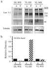
Lan 3-2 epitope upregulation after nerve cord lesion. (a) Lan 3-2 epitope expression was upregulated in segments of ventrally lesioned animals (VL) that generated ectopic heads (EH) or wound healing responses (WH). Western blot analysis of protein extracts from dorsally lesioned animals (DL-WH) revealed protein bands near 210 and 130 kDa and these high-molecular weight protein bands were enriched in VL animals. A 66kDa Lan 3-2 positive protein, a putative molecular marker of neural morphallaxis, was not highly expressed in DL-WH animals, but was enriched in ventrally lesioned animals in segments posterior to the forming ectopic head (VL-EH; column P). The 66kDa band was detectable with the Lan 3-2 antibody both anterior and posterior to the site of wound healing in VL-WH animals (columns A and P). Tubulin antibody was utilized as a loading control. (b) Quantification of the 66kDa morphallaxis marker protein in Western blots was performed with densitometry and was expressed as the ratio of 66kDa band relative to the density of the tubulin loading control band. Densitometry demonstrated that expression of this protein was 7–8-fold greater in segments posterior (P) to the ectopic head of VL-EH animals than in any other protein extract groups. Solid bars indicate densitometry data from extracts of segments anterior (A) to lesion sites and striped bars indicate data from segments posterior (P) to the lesion sites.
Fig. 3
Fig. 3
Segment position-dependent formation of ectopic heads. The dorsal–ventral location and anterior–posterior segmental position of ablation sites (upper illustration) affected the percentage of ectopic heads produced in lesioned animals (lower histogram). Dorsally (D) lesioned worms did not form ectopic heads, irrespective of the segmental position of the ablation. However, ectopic heads produced in ventrally (V) lesioned worms varied depending on the segmental position of the lesion. Specifically, ectopic heads were produced in 71% of worms (n = 23) with ventral ablations at segment 50 (within the zone of giant fiber sensory field overlap; gray area in illustration). This percentage was significantly greater than those found at other ventral lesion sites (**, P<0.05; segment 25, n = 43; segment 75, n = 30; segment 100, n = 55), whether within MGF (black area) or LGF sensory fields (white area of the illustration). The percentage of animals with ectopic heads at ventral lesion sites was significantly greater than corresponding dorsal lesion sites where data were collected (*, P<0.05; nd indicates no data collected).
Fig. 4
Fig. 4
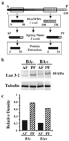
Lan 3-2 epitope upregulation during diminished segmental regeneration. (a) Protein extracts were produced from worm fragments treated with 10mM boric acid for 1 week. As indicated in the illustration, very few new segments formed after boric acid treatment. Boric acid treated zooids and controls incubated in only spring water for 1 week (not shown) were then cultured for an additional 2 weeks earlier to homogenization and protein extraction. Before protein analysis, any newly formed head or tail segments were removed and discarded. A and P designates anterior and posterior. AF and PF indicate anterior and posterior fragments, respectively. Segment numbers are defined below each illustrated animal. (b) Upregulation of the 66kDa Lan 3-2 positive protein was not affected by boric acid treatment (BA+) as compared with Western blots of control fragments (BA−) treated only with spring water. Note that the 66kDa protein band was enriched in posterior fragments (PF), but not in anterior fragments (AF). Tubulin expression was not affected by boric acid and was used as a gel loading control. (c) Quantification of the 66kDa morphallaxis marker protein in Western blots was performed with densitometry and was expressed as the ratio of 66kDa band relative to the density of the tubulin loading control band. Densitometry demonstrated that expression of this protein was 2––3-fold greater in posterior fragments (PF), as compared with anterior fragments (AF), regardless of whether they were treated with boric acid (BA+) or were treated with spring water (BA−).
Fig. 5
Fig. 5
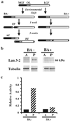
Morphallaxis protein expression persists after block of asexual fission. (a) Animals induced into asexual fission by an environmental shift in temperature were treated with 10 mM boric acid in spring water. Control animals, not treated with boric acid (BA−), formed fission planes (PF) within the region of overlay of MGF and LGF sensory fields (gray area in the illustrations) by 1 week post-shift. The formation of fission planes was accompanied by the emergence of MGF sensory fields (black regions of the illustrations) just posterior to the fission plane. These animals fragmented into two zooids, anterior (AZ) and posterior (PZ), by 3 weeks post-shift. Animals treated with boric acid after the temperature shift (BA+) did not form fission planes and did not fragment. However, analysis of sensory fields using behavioral and electrophysiology assays revealed that weak neural morphallaxis occurred, producing changes in sensory-to-interneuronal connectivity (emergence of ectopic MGF activation; striped region of sensory field map). (b) Lan 3-2 epitope expression was upregulated in segments immediately posterior (P) to the fission site of control animals not treated with boric acid (BA−), but not in segments anterior (A) to the fission site. Although boric acid blocked fission plane formation in treated animals (BA+) and blocked subsequent asexual fragmentation without a significant effect on neural morphallaxis, upregulation of the 66kDa glycoprotein was decreased, but not abolished in segments anterior (A) and posterior (P) to the predicted fission zone. Tubulin expression was also not affected by boric acid treatment and was used as a gel loading control. (c) Quantification of the 66 kDa morphallaxis marker protein in Western blots was performed with densitometry and was expressed as the ratio of 66 kDa band relative to the density of the tubulin loading control band. Densitometry demonstrated that expression of this protein was 6–7-fold greater in segments posterior (P) to the fission site of control animals (BA−), as compared with segments anterior to the fission site (A) in these animals. Levels of this protein were not different between anterior and posterior segments of boric acid treated animals (BA+) and both were many fold lower in their expression of this protein vs. control posterior segment levels. Solid bars indicate densitometry data from extracts of segments anterior (A) to lesion sites and striped bars indicate data from segments posterior (P) to the lesion sites.
Fig. 6
Fig. 6
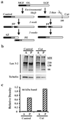
Body transection blocks ongoing asexual fragmentation, not Lan 3-2 epitope upregulation. (a) Asexual reproduction was induced in whole animals using an environmental shift paradigm. The posterior one-third of experimental animals (last 50 segments) was removed 2 days after environmental shift by a body transection at segment number 100. A population of worms that was not amputated and continued to produce fission planes (PF) and fragment asexually into anterior (AZ) and posterior zooids (PZ). Worms that were transected did not complete asexual reproduction. Although fission planes (PF) were obvious at 1 week post-shift, they were absent by 3 weeks post-shift. Changes in giant fiber sensory fields were also reversed in posterior-cut animals by 3 weeks. Black regions of the animal illustrations indicate medial giant fiber (MGF) sensory fields. White regions indicate lateral giant fiber (LGF) sensory fields. The gray area in the illustrations indicates the region of overlap (OL) between MGF and LGF sensory fields. (b) Immunoblot analysis demonstrated similar Lan 3-2 epitope expression profiles in protein extracts from control (asexually reproducing) and cut (aborted asexual reproduction) animals. Protein extracts were created from segments anterior (A) to and posterior (P) to the fission site (48±10). Upregulation of the 66kDa (Lan 3-2 positive) morphallactic protein band was detected in both control and cut animals in segments posterior, but not anterior, to the site of fission plane formation. A tubulin antibody was used as a loading control. (c) Quantification of the 66kDa morphallaxis marker protein in Western blots was performed with densitometry and was expressed as the ratio of 66kDa band relative to the density of the tubulin loading control band. Densitometry demonstrated that expression of this protein was 6–10-fold greater in segments posterior (P) to the fission site of both animals asexually reproducing (Control) and animals whose fission has been aborted owing to body transection (Cut), as compared with segments anterior to fission sites (A) in these groups.

Similar articles

Cited by

References

    1. Achee NL, Zoran MJ. Short-term and long-term alterations in neuronal excitability during injury-induced axonal regeneration in ganglia and cell culture. Invert Neurosci. 1996;2:189–198.
    1. Agata K, Saito Y, Nakajima E. Unifying principles of regeneration I: epimorphosis versus morphallaxis. Dev Growth Differ. 2007;49:73–78. - PubMed
    1. Ambron RT, Walters ET. Priming events and retrograde injury signals. Mol Neurobiol. 1996;13:61–79. - PubMed
    1. Ambron RT, Schmied R, Huang CC, Smedman M. A signal sequence mediates the retrograde transport of proteins from the axon periphery to the cell body and then into the nucleus. J Neurosci. 1992;12:2813–2818. - PMC - PubMed
    1. Bedi SS, Glanzman DL. Axonal rejoining inhibits injury-induced long-term changes in Aplysia sensory neurons in vitro. J Neurosci. 2001;21:9667–9677. - PMC - PubMed

Publication types

MeSH terms

LinkOut - more resources