

Distinct roles of the three Akt isoforms in lactogenic differentiation and involution
- PMID: 18561256
- PMCID: PMC2871282
- DOI: 10.1002/jcp.21518
Distinct roles of the three Akt isoforms in lactogenic differentiation and involution
Abstract
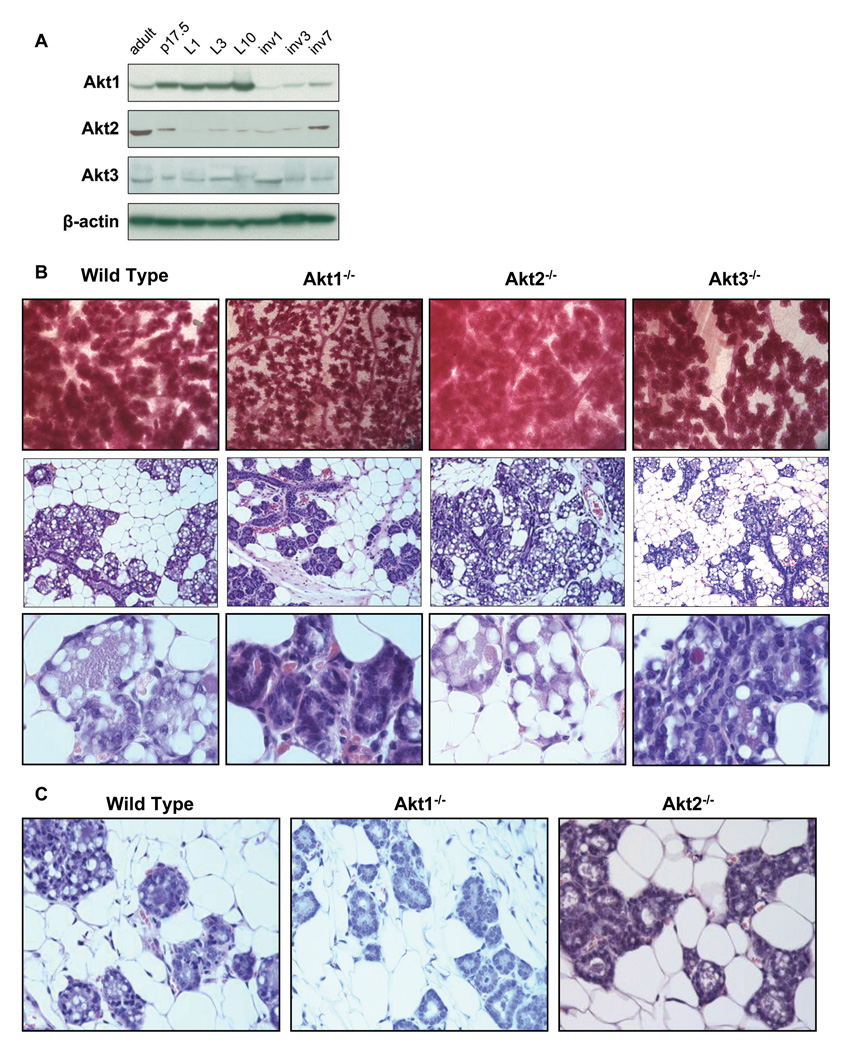
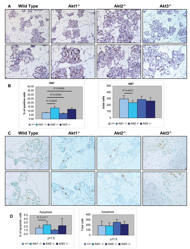
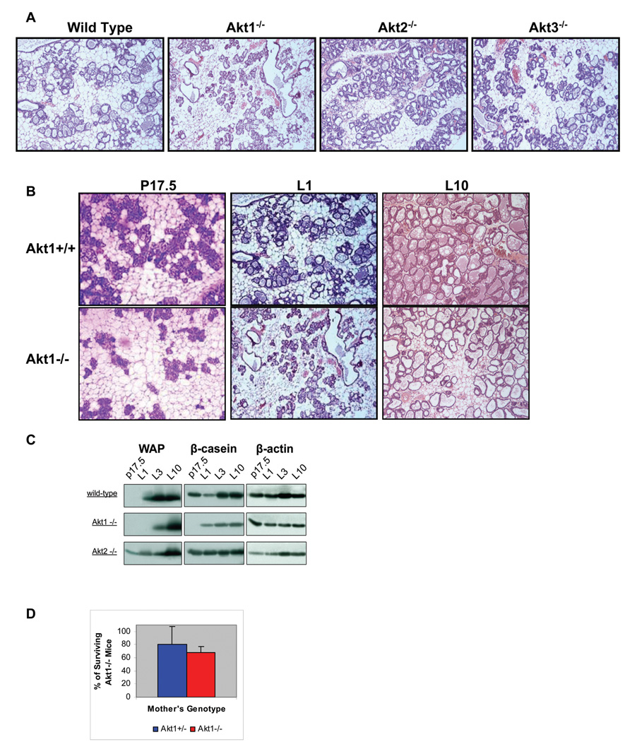
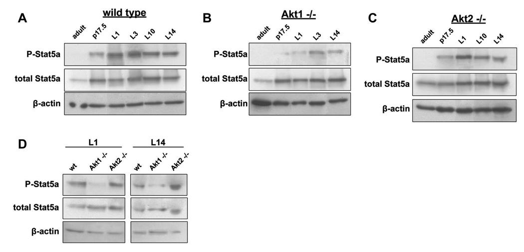
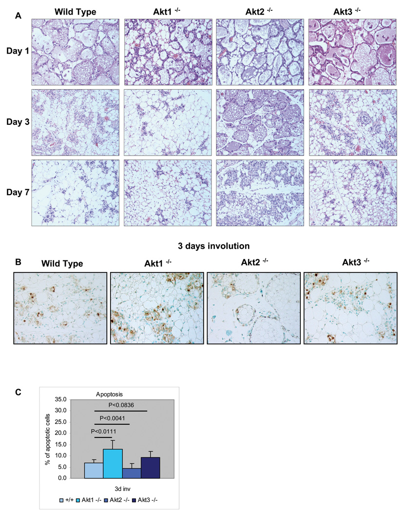
The three Akt isoforms differ in their ability to transduce oncogenic signals initiated by the Neu and PyMT oncogenes in mammary epithelia. As a result, ablation of Akt1 inhibits and ablation of Akt2 accelerates mammary tumor development by both oncogenes, while ablation of Akt3 is phenotypically almost neutral. Since the risk of breast cancer development in humans correlates with multiple late pregnancies, we embarked on a study to determine whether individual Akt isoforms also differ in their ability to transduce hormonal and growth factor signals during pregnancy, lactation and post-lactation involution. The results showed that the ablation of Akt1 delays the differentiation of the mammary epithelia during pregnancy and lactation, and that the ablation of Akt2 has the opposite effect. Finally, ablation of Akt3 results in minor defects, but its phenotype is closer to that of the wild type mice. Whereas the phenotype of the Akt1 ablation is cell autonomous, that of Akt2 is not. The ablation of Akt1 promotes apoptosis and accelerates involution, whereas the ablation of Akt2 inhibits apoptosis and delays involution. Mammary gland differentiation during pregnancy depends on the phosphorylation of Stat5a, which is induced by prolactin, a hormone that generates signals transduced via Akt. Here we show that the ablation of Akt1, but not the ablation of Akt2 or Akt3 interferes with the phosphorylation of Stat5a during late pregnancy and lactation. We conclude that the three Akt isoforms have different roles in mammary gland differentiation during pregnancy and this may reflect differences in hormonal signaling.
(c) 2008 Wiley-Liss, Inc
Figures
Similar articles
-
ErbB3 drives mammary epithelial survival and differentiation during pregnancy and lactation.Breast Cancer Res. 2017 Sep 8;19(1):105. doi: 10.1186/s13058-017-0893-7. Breast Cancer Res. 2017. PMID: 28886748 Free PMC article.
-
Akt is required for Stat5 activation and mammary differentiation.Breast Cancer Res. 2010;12(5):R72. doi: 10.1186/bcr2640. Epub 2010 Sep 17. Breast Cancer Res. 2010. PMID: 20849614 Free PMC article.
-
Akt1 ablation inhibits, whereas Akt2 ablation accelerates, the development of mammary adenocarcinomas in mouse mammary tumor virus (MMTV)-ErbB2/neu and MMTV-polyoma middle T transgenic mice.Cancer Res. 2007 Jan 1;67(1):167-77. doi: 10.1158/0008-5472.CAN-06-3782. Cancer Res. 2007. PMID: 17210696
-
Transcription factor activities and gene expression during mouse mammary gland involution.J Mammary Gland Biol Neoplasia. 1999 Apr;4(2):145-52. doi: 10.1023/a:1018721107061. J Mammary Gland Biol Neoplasia. 1999. PMID: 10426393 Review.
-
Crosstalk between STAT5 activation and PI3K/AKT functions in normal and transformed mammary epithelial cells.Mol Cell Endocrinol. 2017 Aug 15;451:31-39. doi: 10.1016/j.mce.2017.04.025. Epub 2017 May 8. Mol Cell Endocrinol. 2017. PMID: 28495456 Free PMC article. Review.
Cited by
-
Effects of glucose on lactose synthesis in mammary epithelial cells from dairy cow.BMC Vet Res. 2016 May 26;12:81. doi: 10.1186/s12917-016-0704-x. BMC Vet Res. 2016. PMID: 27229304 Free PMC article.
-
Three Novel Players: PTK2B, SYK, and TNFRSF21 Were Identified to Be Involved in the Regulation of Bovine Mastitis Susceptibility via GWAS and Post-transcriptional Analysis.Front Immunol. 2019 Aug 6;10:1579. doi: 10.3389/fimmu.2019.01579. eCollection 2019. Front Immunol. 2019. PMID: 31447828 Free PMC article.
-
Akt1 is essential for postnatal mammary gland development, function, and the expression of Btn1a1.PLoS One. 2011;6(9):e24432. doi: 10.1371/journal.pone.0024432. Epub 2011 Sep 7. PLoS One. 2011. PMID: 21915327 Free PMC article.
-
Differentiation of the mammary epithelial cell during involution: implications for breast cancer.J Mammary Gland Biol Neoplasia. 2009 Jun;14(2):159-70. doi: 10.1007/s10911-009-9121-0. Epub 2009 May 1. J Mammary Gland Biol Neoplasia. 2009. PMID: 19408104 Review.
-
ERBB Receptors and Their Ligands in the Developing Mammary Glands of Different Species: Fifteen Characters in Search of an Author.J Mammary Gland Biol Neoplasia. 2023 May 23;28(1):10. doi: 10.1007/s10911-023-09538-w. J Mammary Gland Biol Neoplasia. 2023. PMID: 37219601 Free PMC article. Review.
References
-
- Bellacosa A, Kumar CC, Di Cristofano A, Testa JR. Activation of AKT kinases in cancer: implications for therapeutic targeting. Adv Cancer Res. 2005;94:29–86. - PubMed
-
- Bellacosa A, Testa JR, Staal SP, Tsichlis PN. A retroviral oncogene, akt, encoding a serine-threonine kinase containing an SH2-like region. Science. 1991;254(5029):274–277. - PubMed
-
- Bowe DB, Kenney NJ, Adereth Y, Maroulakou IG. Suppression of Neu-induced mammary tumor growth in cyclin D1 deficient mice is compensated for by cyclin E. Oncogene. 2002;21(2):291–298. - PubMed
-
- Boxer RB, Stairs DB, Dugan KD, Notarfrancesco KL, Portocarrero CP, Keister BA, Belka GK, Cho H, Rathmell JC, Thompson CB, Birnbaum MJ, Chodosh LA. Isoform-specific requirement for Akt1 in the developmental regulation of cellular metabolism during lactation. Cell Metab. 2006;4(6):475–490. - PubMed
-
- Brisken C, Kaur S, Chavarria TE, Binart N, Sutherland RL, Weinberg RA, Kelly PA, Ormandy CJ. Prolactin controls mammary gland development via direct and indirect mechanisms. Dev Biol. 1999;210(1):96–106. - PubMed
Publication types
MeSH terms
Substances
Grants and funding
LinkOut - more resources
Full Text Sources
Research Materials
Miscellaneous
