

Dedicated breast computed tomography: volume image denoising via a partial-diffusion equation based technique
- PMID: 18561671
- PMCID: PMC2809732
- DOI: 10.1118/1.2903436
Dedicated breast computed tomography: volume image denoising via a partial-diffusion equation based technique
Abstract
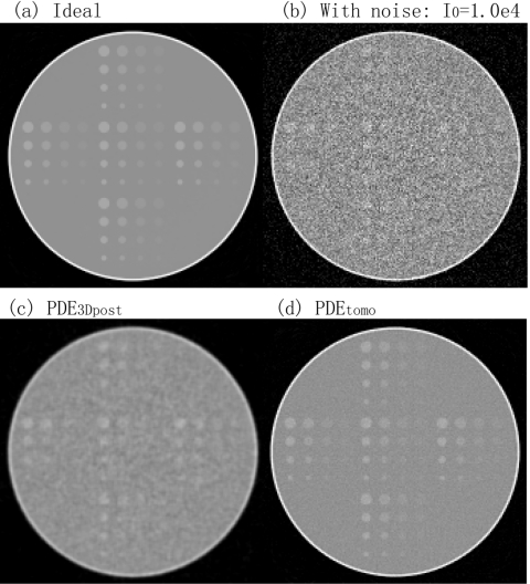
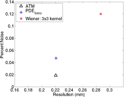
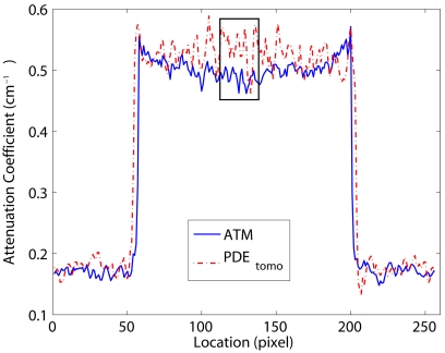
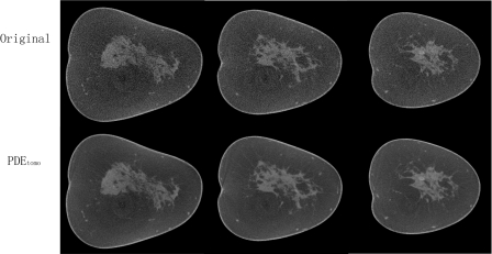
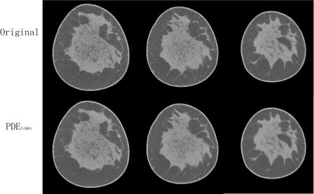
Dedicated breast computed tomography (CT) imaging possesses the potential for improved lesion detection over conventional mammograms, especially for women with dense breasts. The breast CT images are acquired with a glandular dose comparable to that of standard two-view mammography for a single breast. Due to dose constraints, the reconstructed volume has a non-negligible quantum noise when thin section CT slices are visualized. It is thus desirable to reduce noise in the reconstructed breast volume without loss of spatial resolution. In this study, partial diffusion equation (PDE) based denoising techniques specifically for breast CT were applied at different steps along the reconstruction process and it was found that denoising performed better when applied to the projection data rather than reconstructed data. Simulation results from the contrast detail phantom show that the PDE technique outperforms Wiener denoising as well as adaptive trimmed mean filter. The PDE technique increases its performance advantage relative to Wiener techniques when the photon fluence is reduced. With the PDE technique, the sensitivity for lesion detection using the contrast detail phantom drops by less than 7% when the dose is cut down to 40% of the two-view mammography. For subjective evaluation, the PDE technique was applied to two human subject breast data sets acquired on a prototype breast CT system. The denoised images had appealing visual characteristics with much lower noise levels and improved tissue textures while maintaining sharpness of the original reconstructed volume.
Figures
References
-
- ACS, American Cancer Society: Cancer Facts and Figures 2008 (American Cancer Society, Atlanta, Georgia, 2008).
-
- Jackson V. P., Hendrick R. E., Feig S. A., and Kopans D. B., “Imaging of the radiographically dense breast,” Radiology 188, 297–301 (1993). - PubMed
-
- Niklason L. T. et al., “Digital tomosynthesis in breast imaging,” Radiology 205, 399–406 (1997). - PubMed
Publication types
MeSH terms
Grants and funding
LinkOut - more resources
Full Text Sources
Other Literature Sources
Medical
