

Imaging performance of an amorphous selenium digital mammography detector in a breast tomosynthesis system
- PMID: 18561674
- PMCID: PMC2673645
- DOI: 10.1118/1.2903425
Imaging performance of an amorphous selenium digital mammography detector in a breast tomosynthesis system
Abstract
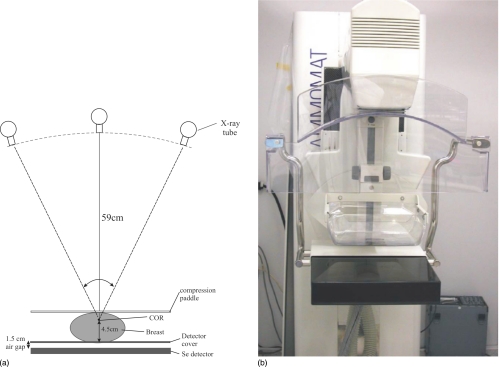
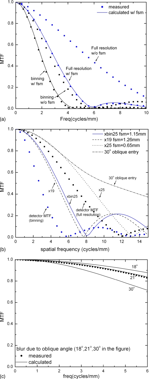
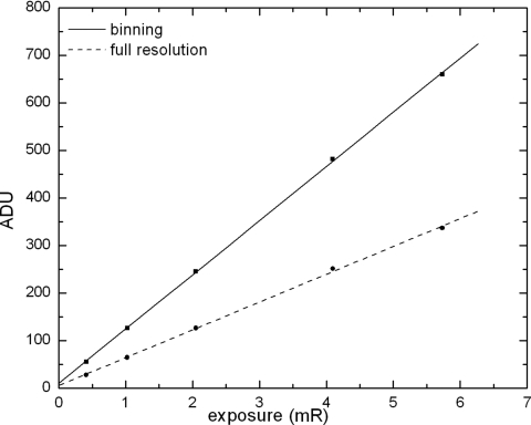
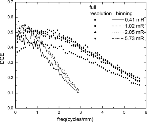
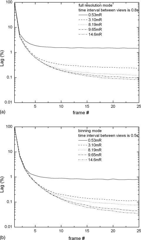
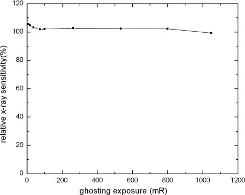
In breast tomosynthesis a rapid sequence of N images is acquired when the x-ray tube sweeps through different angular views with respect to the breast. Since the total dose to the breast is kept the same as that in regular mammography, the exposure used for each image of tomosynthesis is 1/N. The low dose and high frame rate pose a tremendous challenge to the imaging performance of digital mammography detectors. The purpose of the present work is to investigate the detector performance in different operational modes designed for tomosynthesis acquisition, e.g., binning or full resolution readout, the range of view angles, and the number of views N. A prototype breast tomosynthesis system with a nominal angular range of +/-25 degrees was used in our investigation. The system was equipped with an amorphous selenium (a-Se) full field digital mammography detector with pixel size of 85 microm. The detector can be read out in full resolution or 2 x 1 binning (binning in the tube travel direction). The focal spot blur due to continuous tube travel was measured for different acquisition geometries, and it was found that pixel binning, instead of focal spot blur, dominates the detector modulation transfer function (MTF). The noise power spectrum (NPS) and detective quantum efficiency (DQE) of the detector were measured with the exposure range of 0.4-6 mR, which is relevant to the low dose used in tomosynthesis. It was found that DQE at 0.4 mR is only 20% less than that at highest exposure for both detector readout modes. The detector temporal performance was categorized as lag and ghosting, both of which were measured as a function of x-ray exposure. The first frame lags were 8% and 4%, respectively, for binning and full resolution mode. Ghosting is negligible and independent of the frame rate. The results showed that the detector performance is x-ray quantum noise limited at the low exposures used in each view of tomosynthesis, and the temporal performance at high frame rate (up to 2 frames per second) is adequate for tomosynthesis.
Figures

Similar articles
-
Experimental validation of a three-dimensional linear system model for breast tomosynthesis.Med Phys. 2009 Jan;36(1):240-51. doi: 10.1118/1.3040178. Med Phys. 2009. PMID: 19235392 Free PMC article.
-
Three-dimensional linear system analysis for breast tomosynthesis.Med Phys. 2008 Dec;35(12):5219-32. doi: 10.1118/1.2996014. Med Phys. 2008. PMID: 19175081 Free PMC article.
-
A computer simulation platform for the optimization of a breast tomosynthesis system.Med Phys. 2007 Mar;34(3):1098-109. doi: 10.1118/1.2558160. Med Phys. 2007. PMID: 17441255
-
Digital tomosynthesis: technique.Radiol Clin North Am. 2014 May;52(3):489-97. doi: 10.1016/j.rcl.2014.01.003. Radiol Clin North Am. 2014. PMID: 24792651 Review.
-
Direct-conversion flat-panel imager with avalanche gain: feasibility investigation for HARP-AMFPI.Med Phys. 2008 Dec;35(12):5207-18. doi: 10.1118/1.3002314. Med Phys. 2008. PMID: 19175080 Free PMC article. Review.
Cited by
-
Toward Scintillator High-Gain Avalanche Rushing Photoconductor Active Matrix Flat Panel Imager (SHARP-AMFPI): Initial fabrication and characterization.Med Phys. 2018 Feb;45(2):794-802. doi: 10.1002/mp.12693. Epub 2017 Dec 18. Med Phys. 2018. PMID: 29171067 Free PMC article.
-
Novel method to determine recursive filtration and noise reduction in fluoroscopic imaging - a comparison of four different vendors.J Appl Clin Med Phys. 2021 Jan;22(1):281-292. doi: 10.1002/acm2.13115. Epub 2020 Dec 14. J Appl Clin Med Phys. 2021. PMID: 33315295 Free PMC article.
-
Experimental validation of a three-dimensional linear system model for breast tomosynthesis.Med Phys. 2009 Jan;36(1):240-51. doi: 10.1118/1.3040178. Med Phys. 2009. PMID: 19235392 Free PMC article.
-
Carbon nanotube electron field emitters for x-ray imaging of human breast cancer.Nanotechnology. 2014 Jun 20;25(24):245704. doi: 10.1088/0957-4484/25/24/245704. Epub 2014 May 28. Nanotechnology. 2014. PMID: 24869902 Free PMC article.
-
Digital breast tomosynthesis: studies of the effects of acquisition geometry on contrast-to-noise ratio and observer preference of low-contrast objects in breast phantom images.Phys Med Biol. 2014 Oct 7;59(19):5883-902. doi: 10.1088/0031-9155/59/19/5883. Epub 2014 Sep 11. Phys Med Biol. 2014. PMID: 25211509 Free PMC article.
References
-
- Niklason L. T. et al. , “Digital tomosynthesis in breast imaging,” Radiology 205, 399–406 (1997). - PubMed
-
- Smith A., “Full-field breast tomosynthesis,” Radiol. Manage. 27, 25–31 (2005). - PubMed
-
- Chen Y., Lo J. Y., and J. T.DobbinsIII, “Impulse response analysis for several digital tomosynthesis mammography reconstruction algorithms,” Proc. SPIE PSISDG10.1117/12.595684 5745, 541–549 (2005). - DOI
Publication types
MeSH terms
Substances
Grants and funding
LinkOut - more resources
Full Text Sources
Other Literature Sources
Medical
Miscellaneous
