

The apparent critical isotherm for cryoinsult-induced osteonecrotic lesions in emu femoral heads
- PMID: 18561937
- PMCID: PMC2612542
- DOI: 10.1016/j.jbiomech.2008.04.032
The apparent critical isotherm for cryoinsult-induced osteonecrotic lesions in emu femoral heads
Abstract
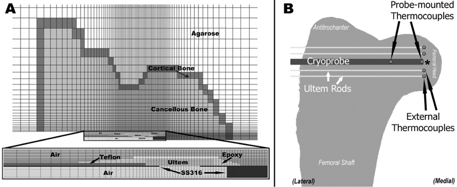
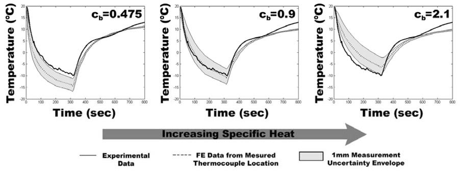
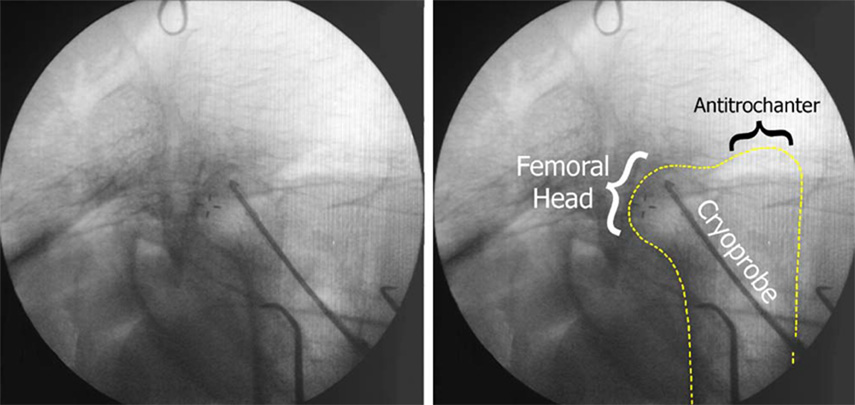
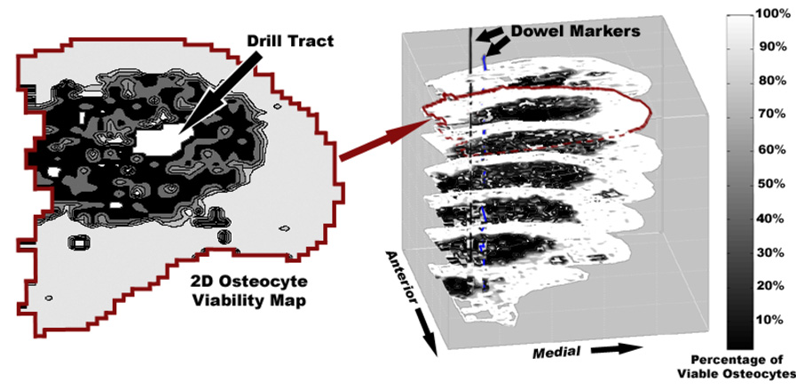
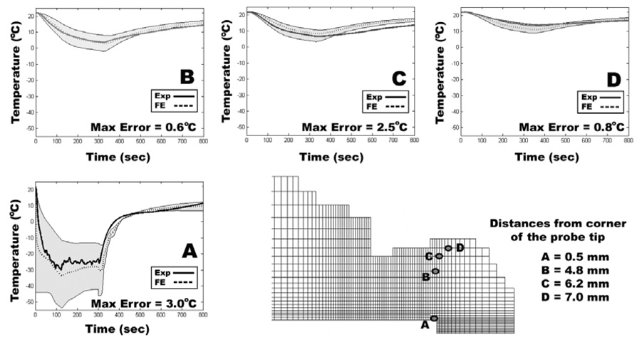
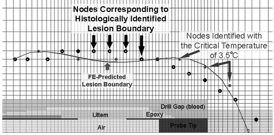
Cryoinsult-induced osteonecrosis (ON) in the emu femoral head provides a unique opportunity to systematically explore the pathogenesis of ON in an animal model that progresses to human-like femoral head collapse. Among the various characteristics of cryoinsult, the maximally cold temperature attained is one plausible determinant of tissue necrosis. To identify the critical isotherm required to induce development of ON in the cancellous bone of the emu femoral head, a thermal finite element (FE) model of intraoperative cryoinsults was developed. Thermal material property values of emu cancellous bone were estimated from FE simulations of cryoinsult to emu cadaver femora, by varying model properties until the FE-generated temperatures matched corresponding thermocouple measurements. The resulting FE model, with emu bone-specific thermal properties augmented to include blood flow effects, was then used to study intraoperatively performed in vivo cryoinsults. Comparisons of minimum temperatures attained at FE nodes corresponding to the three-dimensional histologically apparent boundary of the region of ON were made for six experimental cryoinsults. Series-wide, a critical isotherm of 3.5 degrees C best corresponded to the boundary of the osteonecrotic lesions.
Figures
Similar articles
-
Cryoinsult parameter effects on the histologically apparent volume of experimentally induced osteonecrotic lesions.J Orthop Res. 2011 Jun;29(6):931-7. doi: 10.1002/jor.21342. Epub 2011 Jan 21. J Orthop Res. 2011. PMID: 21259339 Free PMC article.
-
Emu model of full-range femoral head osteonecrosis induced focally by an alternating freezing and heating insult.J Int Med Res. 2011;39(1):187-98. doi: 10.1177/147323001103900120. J Int Med Res. 2011. PMID: 21672321
-
A new animal model of femoral head osteonecrosis: one that progresses to human-like mechanical failure.J Orthop Res. 2002 Mar;20(2):303-9. doi: 10.1016/S0736-0266(01)00108-5. J Orthop Res. 2002. PMID: 11918310
-
Osteonecrosis of the Femoral Head: an Updated Review of ARCO on Pathogenesis, Staging and Treatment.J Korean Med Sci. 2021 Jun 21;36(24):e177. doi: 10.3346/jkms.2021.36.e177. J Korean Med Sci. 2021. PMID: 34155839 Free PMC article. Review.
-
Application of biomaterials in treating early osteonecrosis of the femoral head: Research progress and future perspectives.Acta Biomater. 2023 Jul 1;164:15-73. doi: 10.1016/j.actbio.2023.04.005. Epub 2023 Apr 18. Acta Biomater. 2023. PMID: 37080444 Review.
Cited by
-
Temperature Threshold Values of Bone Necrosis for Thermo-Explantation of Dental Implants-A Systematic Review on Preclinical In Vivo Research.Materials (Basel). 2020 Aug 6;13(16):3461. doi: 10.3390/ma13163461. Materials (Basel). 2020. PMID: 32781597 Free PMC article. Review.
-
A pilot cadaveric study of temperature and adjacent tissue changes after exposure of magnetic-controlled growing rods to MRI.Eur Spine J. 2017 Jun;26(6):1618-1623. doi: 10.1007/s00586-016-4918-1. Epub 2017 Jan 9. Eur Spine J. 2017. PMID: 28070684
-
Heat Generation During Initial Osteotomy for Implant Site Preparation: An In Vitro Measurement Study.J Maxillofac Oral Surg. 2023 Jun;22(2):313-320. doi: 10.1007/s12663-022-01800-8. Epub 2022 Oct 27. J Maxillofac Oral Surg. 2023. PMID: 37122802 Free PMC article.
-
A simple method for establishing an ostrich model of femoral head osteonecrosis and collapse.J Orthop Surg Res. 2015 May 21;10:74. doi: 10.1186/s13018-015-0218-4. J Orthop Surg Res. 2015. PMID: 25994205 Free PMC article.
-
A canine model of femoral head osteonecrosis induced by an ethanol injection navigated by a novel template.Int J Med Sci. 2013 Aug 27;10(11):1451-8. doi: 10.7150/ijms.6314. eCollection 2013. Int J Med Sci. 2013. PMID: 24046517 Free PMC article.
References
-
- Auge BK, Santa-Cruz RW, Polascik TJ. Effect of freeze time during renal cryoablation: a swine model. Journal of Endourology. 2006;20(12):1101–1105. - PubMed
-
- Baust J, Gage AA, Ma H, Zhang CM. Minimally invasive cryosurgery--technological advances. Cryobiology. 1997;34(4):373–384. - PubMed
-
- Baust JG, Gage AA. The molecular basis of cryosurgery. BJU International. 2005;95(9):1187–1191. - PubMed
-
- Biyikli S, Modest MF, Tarr R. Measurements of thermal properties for human femora. Journal of Biomedical Materials Research. 1986;20(9):1335–1345. - PubMed
-
- Clattenburg R, Cohen J, Conner S, Cook N. Thermal properties of cancellous bone. Journal of Biomedical Materials Research. 1975;9(2):169–182. - PubMed
Publication types
MeSH terms
Grants and funding
LinkOut - more resources
Full Text Sources
Medical
Miscellaneous
