. 2008 Jul 18;380(4):608-22.
doi: 10.1016/j.jmb.2007.11.093. Epub 2007 Dec 5.

Solution structure of the c-terminal dimerization domain of SARS coronavirus nucleocapsid protein solved by the SAIL-NMR method

Affiliations

Solution structure of the c-terminal dimerization domain of SARS coronavirus nucleocapsid protein solved by the SAIL-NMR method

Mitsuhiro Takeda et al. J Mol Biol. .

Abstract

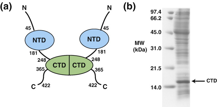
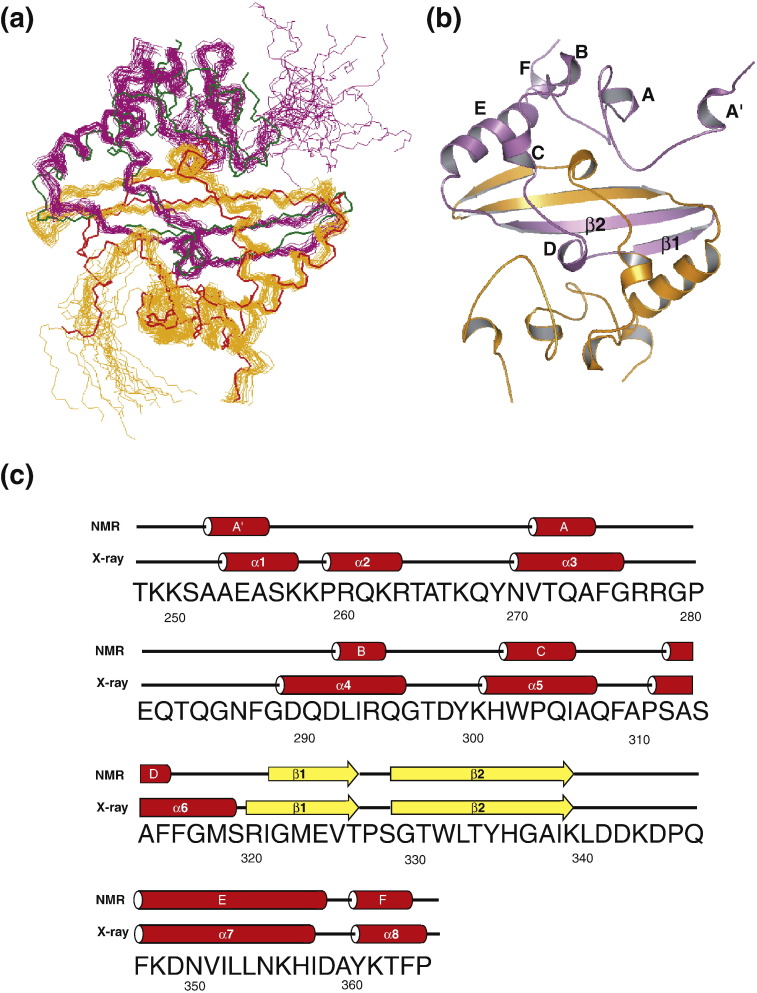
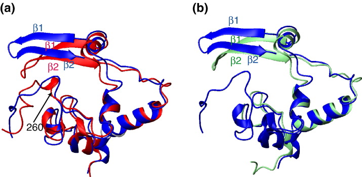
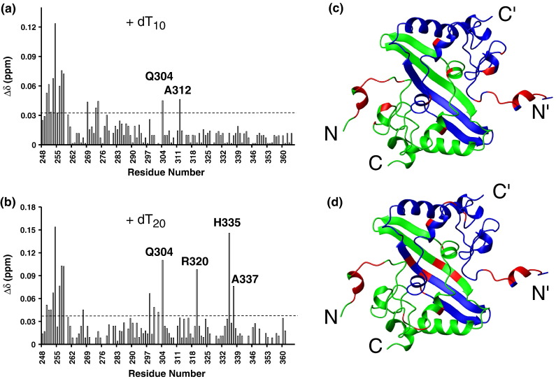
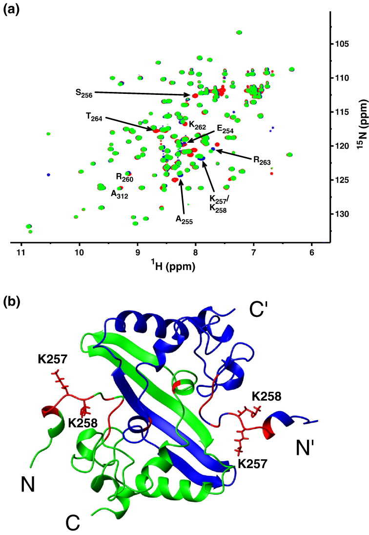
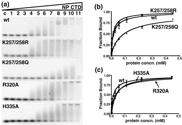
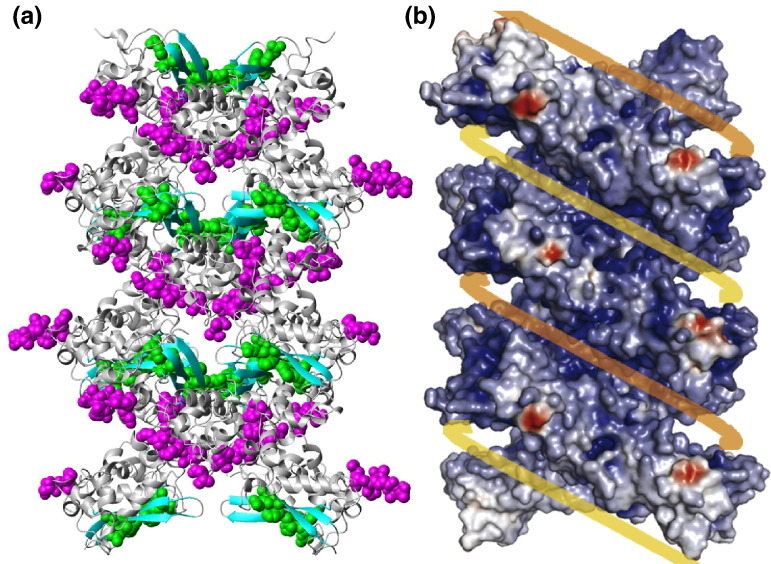
The C-terminal domain (CTD) of the severe acute respiratory syndrome coronavirus (SARS-CoV) nucleocapsid protein (NP) contains a potential RNA-binding region in its N-terminal portion and also serves as a dimerization domain by forming a homodimer with a molecular mass of 28 kDa. So far, the structure determination of the SARS-CoV NP CTD in solution has been impeded by the poor quality of NMR spectra, especially for aromatic resonances. We have recently developed the stereo-array isotope labeling (SAIL) method to overcome the size problem of NMR structure determination by utilizing a protein exclusively composed of stereo- and regio-specifically isotope-labeled amino acids. Here, we employed the SAIL method to determine the high-quality solution structure of the SARS-CoV NP CTD by NMR. The SAIL protein yielded less crowded and better resolved spectra than uniform (13)C and (15)N labeling, and enabled the homodimeric solution structure of this protein to be determined. The NMR structure is almost identical with the previously solved crystal structure, except for a disordered putative RNA-binding domain at the N-terminus. Studies of the chemical shift perturbations caused by the binding of single-stranded DNA and mutational analyses have identified the disordered region at the N-termini as the prime site for nucleic acid binding. In addition, residues in the beta-sheet region also showed significant perturbations. Mapping of the locations of these residues onto the helical model observed in the crystal revealed that these two regions are parts of the interior lining of the positively charged helical groove, supporting the hypothesis that the helical oligomer may form in solution.


Figures

Fig. 1
Fig. 1
Preparation of the SARS-CoV NP CTD. (a) Schematic diagram of the domain architecture of the SARS-CoV NP. (b) SDS-PAGE of the cell-free reaction mixture for the SARS-CoV NP CTD. The left lane shows the molecular weight markers. The band corresponding to the monomer of the SARS-CoV NP CTD is labeled with an arrow.
Fig. 2
Fig. 2
Comparisons of NMR spectra for the SARS-CoV NP CTD between UL and SAIL in the methylene region. Aliphatic region of 1H–13C CT-HSQC for UL (a) and SAIL (b) SARS-CoV NP CTD. Both spectra were acquired under the same conditions. The sample concentration was 0.5 mM. In (b), assignments for the SAIL sample are labeled. (c) Cross-sections from (a) (red) and (b) (black). The peak scales are identical between the UL and SAIL spectra.
Fig. 3
Fig. 3
Comparisons of NMR spectra for the SARS-CoV NP CTD between UL and SAIL in the aromatic region. Chemical structures of the aromatic rings for UL (a) and SAIL (c) phenylalanine. (b and d) Phenylalanine signals of 1H–13C HSQC for UL (b) and SAIL (d) SARS-CoV NP CTD. (e and f) Tyrosine signals of 1H–13C HSQC for UL (e) and SAIL (f) SARS-CoV NP CTD. To demonstrate the absence of the 1Jcc coupling of aromatic rings for SAIL phenylalanine and tyrosine residues, all 1H–13C HSQC spectra for the aromatic regions were recorded without the CT technique.
Fig. 4
Fig. 4
NMR structure of the SARS-CoV NP CTD. (a) Superposition of the 20 lowest-energy NMR structures of the SARS-CoV NP CTD and the corresponding crystal structure spanning residues 248–365. The two subunits in each structure are in orange and magenta for the NMR structure, and in red and green for the crystal structure. (b) Ribbon diagram of the solution structure of the SARS-CoV NP CTD. Secondary structure elements are labeled for one subunit. (c) Sequence of the SARS-CoV NP CTD. The secondary structures for the NMR structure and for the crystal structure spanning residues 248–365 are shown above the sequence, with red cylinders for α-helices and yellow arrows for β-strands.
Fig. 5
Fig. 5
Superposition of the NMR and crystal structures of a CTD monomer of SARS-CoV NP. The mean NMR structure of a CTD monomer (blue) is superposed on the corresponding crystal structure spanning residues 248–365 (a) (red; PDB code 2cjr) and on that encompassing residues 270–370 (b) (light green; PDB code 2gib). In (a), the two structures are superposed on the regions of residues 260–319 and 333–358, where the backbone RMSD between them is 1.45 Å. In (b), the two structures are superposed on the regions of residues 274–319 and 333–358, where the backbone RMSD is 1.26 Å.
Fig. 6
Fig. 6
CSD of SARS-CoV NP CTD titrated with poly-dT ssDNA. Variation of the CSD of SARS-CoV NP CTD titrated with dT10 (a) or dT20 (b). The dashed lines in (a) and (b) represent the cutoff for significant displacements. (c) Spatial locations of residues (red) with CSD values larger than the cutoff value upon titration with dT10 (c) or dT20 (d). The two monomers are in green and blue, respectively.
Fig. 7
Fig. 7
Structure perturbation of K257/K258 double mutants. (a) Overlay of 15N-edited HSQC spectra from wild-type CTD (blue), K257R/K258R (green), and K257Q/K258Q (red) double mutants. Affected resonances are identified by their respective residue types and numbers in the wild-type protein. These are mapped onto the ribbon structure of the CTD dimer in (b).
Fig. 8
Fig. 8
EMSA of SARS-CoV NP CTD mutants. (a) Mobility shift of dT20 bound to wild-type (wt), K257R/K258R (K257/258R), K257Q/K258Q (K257/258Q), R320A, and H335A mutant proteins. The protein concentration was increased by a factor of 2, starting from lane 1 (439 nM) to lane 11 (0.45 mM). Lane C, negative control. (b) Binding curve of the K257/K258 double mutant towards dT20, compared to that of the wild-type protein. (c) Binding curve of the R320A and H335A mutants towards dT20, compared to that of the wild-type protein. Each curve in (b) and (c) represents the best fit from three independent assays. Results are summarized in Table 2.
Fig. 9
Fig. 9
Structure perturbation of R320A and H335A mutants. (a) Overlay of 15N-edited HSQC spectra from the wild-type CTD (blue) and the R320A mutant (red). Affected resonances are identified by their respective residue types and numbers in the wild-type protein. (b) Same as in (a), but with the wild-type CTD (blue) and the H335A mutant (magenta). (c) Mapping of residues affected by the R320A mutation (red) in the solution structure of the SARS-CoV NP CTD dimer. The side chains of R320 are shown in a neon representation. (d) Same as in (c), but showing the residues affected by the H335A mutation (in magenta). The side chain of H335 is also shown.
Fig. 10
Fig. 10
Spatial locations of the nucleic-acid-binding sites in the helical packing model of the SARS-CoV NP CTD crystal. (a) Binding sites are shown in CPK models, with the N-termini residues in magenta and with the residues on the β-sheet (R320, H335, and A337) in green. The rest of the molecules are shown in a gray ribbon representation, except for the β-sheets, which are in cyan. (b) Surface charge representation of the proposed helical supramolecular complex (adapted from Chen et al.5). The yellow and orange lines represent viral RNA strands. Notice that the binding sites in (a) are located in the positively charged grooves within the supramolecular complex.

Similar articles

Cited by

References

    1. Drosten C., Gunther S., Preiser W., van der Werf S., Brodt H.R., Becker S., et al. Identification of a novel coronavirus in patients with severe acute respiratory syndrome. N. Engl. J. Med. 2003;348:1967–1976. - PubMed
    1. Ksiazek T., Erdman D., Goldsmith C., Zaki S., Peret T., Emery S., et al. A novel coronavirus associated with severe acute respiratory syndrome. N. Engl. J. Med. 2003;348:1953–1966. - PubMed
    1. Chang C., Sue S., Yu T., Hsieh C., Tsai C., Chiang Y., et al. Modular organization of SARS coronavirus nucleocapsid protein. Biomed. Sci. 2005;13:59–72. - PMC - PubMed
    1. Huang Q., Yu L., Petros A.M., Gunasekera A., Liu Z., Xu N., et al. Structure of the N-terminal RNA-binding domain of the SARS CoV nucleocapsid protein. Biochemistry. 2004;43:6059–6063. - PubMed
    1. Chen C., Chang C., Chang Y., Sue S., Bai H., Riang L., et al. Structure of the SARS coronavirus nucleocapsid protein RNA-binding dimerization domain suggests a mechanism for helical packaging of viral RNA. J. Mol. Biol. 2007;368:1075–1086. - PMC - PubMed

Publication types

MeSH terms

Substances

Associated data

LinkOut - more resources