. 2008 Aug 13;199(1-2):46-55.
doi: 10.1016/j.jneuroim.2008.05.003. Epub 2008 Jun 17.

Lipoic acid stimulates cAMP production via the EP2 and EP4 prostanoid receptors and inhibits IFN gamma synthesis and cellular cytotoxicity in NK cells

Affiliations

Lipoic acid stimulates cAMP production via the EP2 and EP4 prostanoid receptors and inhibits IFN gamma synthesis and cellular cytotoxicity in NK cells

Sonemany Salinthone et al. J Neuroimmunol. .

Abstract

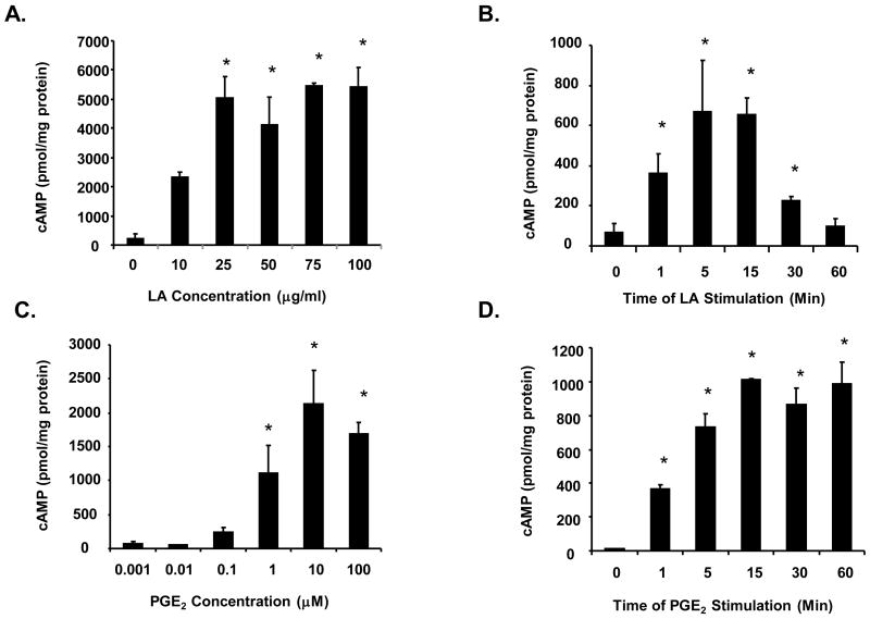
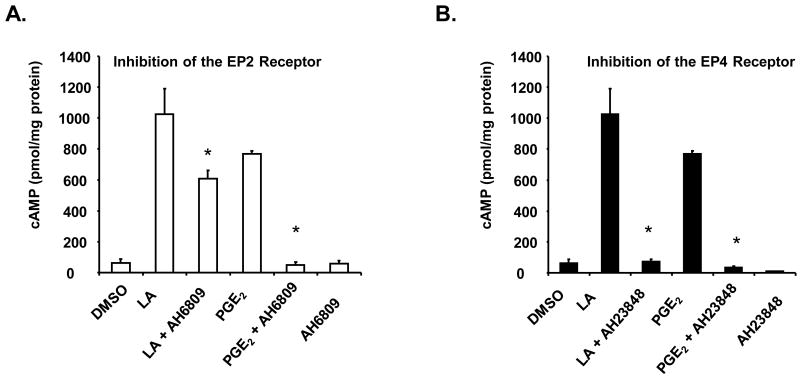
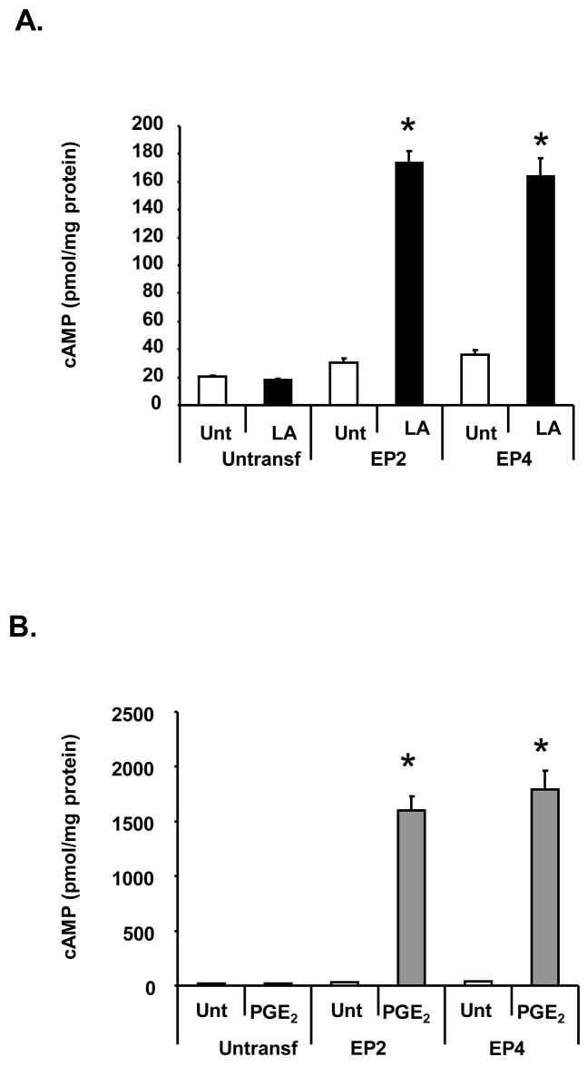
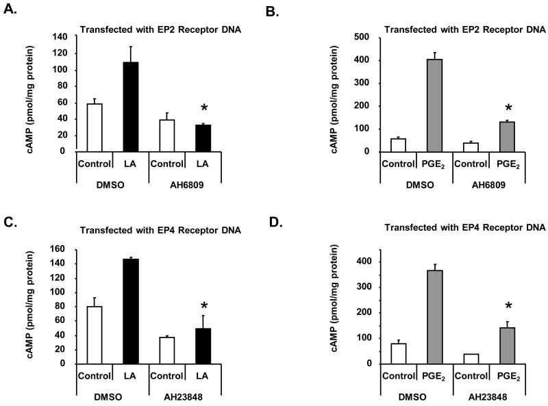
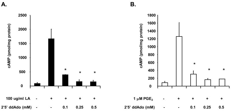
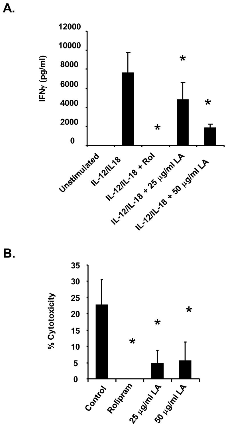
The antioxidant lipoic acid (LA) treats and prevents the animal model of multiple sclerosis (MS), experimental autoimmune encephalomyelitis (EAE). In an effort to understand the therapeutic potential of LA in MS, we sought to define the cellular mechanisms that mediate the effects of LA on human natural killer (NK) cells, which are important in innate immunity as the first line of defense against invading pathogens and tumor cells. We discovered that LA stimulates cAMP production in NK cells in a dose-dependent manner. Studies using pharmacological inhibitors and receptor transfection experiments indicate that LA stimulates cAMP production via activation of the EP2 and EP4 prostanoid receptors and adenylyl cyclase. In addition, LA suppressed interleukin (IL)-12/IL-18 induced IFNgamma secretion and cytotoxicity in NK cells. These novel findings suggest that LA may inhibit NK cell function via the cAMP signaling pathway.


Figures

Figure 1
Figure 1. Lipoic acid and PGE2 stimulates cAMP in a concentration-dependent manner
(A) Purified human NK cells (1–2 × 10⁵) were stimulated with 0, 10, 25, 50, 75 and 100 μg/ml LA for 1 minute and centrifuged. The pellets were resuspended in 0.1 M HCl and lysed with boiling for 10 minutes. The supernatants were used in cAMP assays. N = 4 donors in duplicate, * indicates statistically significant compared to unstimulated control, p < 0.05. (B) Purified human NK cells were stimulated with 100 μg/ml LA for 0, 1, 5, 15, 30 and 60 minutes (B), 0.001, 0.01, 0.1, 1, 10 and 100 μM PGE2 for 1 min (C), or 10 μM PGE2 for 0, 1, 5, 15, 30 and 60 minutes (D). Samples were processed as described in A. N = 3 donors in duplicate for B–D, * indicates statistically significant compared to unstimulated control, p < 0.05.
Figure 2
Figure 2. LA stimulates cAMP production via the EP2 and EP4 receptors
NK cells (1 × 10⁵) were pre-incubated with 50 μM AH6809 (A) or AH23848 (B) for 30 minutes in RPMI and then stimulated with 10 μg/ml LA or 1 μM PGE2 for 1 minute. Media was removed and cells were lysed with 0.1 M HCl and boiling for 10 minutes. cAMP assays were determined by ELISA as described in materials and method. Data are graphical representatives of 3 donors in duplicate. * indicates statistically significant compared to stimulated controls, p< 0.05.
Figure 3
Figure 3. LA stimulates cAMP production in HEK 293 EBNA cells transfected with the EP2 or EP4 receptors
HEK 293 EBNA cells were plated on a 12 well plate at 2 × 10⁵ cells per well for 24 hours. Cells were transfected with 5 μg EP2 or EP4 DNA using lipofectamine 2000 and stimulated 48 hours later with 100 μ/ml LA (A) or 1 nM PGE2 (B) in the presence of 100 μM IBMX for 1 hour. Cells were scraped in 0.1 M HCl and boiled for 10 minutes. The supernatant was then used for determining cAMP levels by ELISA. Depicted is a representative of 3 donors in duplicate. * indicates statistically significant compared to untreated, transfected cells, p< 0.05.
Figure 4
Figure 4. EP2 and EP4 inhibitors block cAMP production in HEK EBNA cells transfected with the EP2 or EP4 receptors
HEK 293 EBNA cells were plated on a 12 well plate at 2 × 10⁵ cells per well for 24 hours. Cells were transfected using lipofectamine 2000 and pre-incubated with AH6809 (A-B) or AH23848 (C-D) 48 hours later in the presence of 100 μM IBMX. After 30 minutes, cells were then stimulated with 100 μg/ml LA (A,C) or 1 nM PGE2 (B,D) for 1 hour. Cells were scraped in 0.1 M HCl and boiled for 10 minutes. The supernatant was then used for determining cAMP levels by ELISA. Depicted is a representative of 3 donors in duplicate. * indicates statistically significant compared to stimulated controls, p< 0.05.
Figure 5
Figure 5. The AC inhibitor (2′5′-dideoxyadenosine or ddAdo) blocks cAMP production in human NK cells
NK cells (1 × 10⁵) were pre-incubated with the indicated concentrations of 2′5′ddAdo for 30 minutes in RPMI and then stimulated with 100 μg/ml LA (A) or 1 μM PGE2 (B) for 1 minute. Media was removed and cells were lysed with 0.1 M HCl and boiling for 10 minutes. cAMP assays were determined by ELISA. Depicted is a representative of 3 donors in duplicate. * indicates statistically significant compared to stimulated controls, p< 0.05.
Figure 6
Figure 6. LA inhibits IFNγ secretion and NK cell cytotoxicity
(A) Purified NK cells were plated on a 96-well plate at 1 × 10⁵ cells per well in RPMI. Cells were pre-treated with 1 mM rolipram (30 min) or 25 or 50 μg/ml LA (1 min) then stimulated with a combination of IL-12/IL-18 for 24 hours. Ten percent FBS was added after treatment to ensure cell survival overnight. Supernatants were used to measure IFNγ secretion by ELISA. Depicted is a representative of 3 donors in duplicate. * indicates statistically significant compared to stimulated control, p< 0.05. (B) LDH release. NK cells were pre-incubated with rolipram or LA prior to addition of K562 target cells at a 6:1 effector:target ratio. The supernatant was removed and used for measurement of LDH release as in materials and method. Depicted is a representative of 3 donors in duplicate. * indicates statistically significant compared to untreated control, p< 0.05.

Similar articles

Cited by

References

    1. Anderson KA, Ribar TJ, Illario M, Means AR. Defective survival and activation of thymocytes in transgenic mice expressing a catalytically inactive form of Ca2+/calmodulin-dependent protein kinase IV. Mol Endocrinol. 1997;11:725–737. - PubMed
    1. Antel JP, McCrea E, Ladiwala U, Qin YF, Becher B. Non-MHC-restricted cell-mediated lysis of human oligodendrocytes in vitro: relation with CD56 expression. J Immunol. 1998;160:1606–1611. - PubMed
    1. Aranami T, Miyake S, Yamamura T. Differential expression of CD11c by peripheral blood NK cells reflects temporal activity of multiple sclerosis. J Immunol. 2006;177:5659–5667. - PubMed
    1. Artwohl M, Muth K, Kosulin K, de Martin R, Holzenbein T, Rainer G, Freudenthaler A, Huttary N, Schmetterer L, Waldhausl WK, Baumgartner-Parzer SM. R-(+)-alpha-lipoic acid inhibits endothelial cell apoptosis and proliferation: involvement of Akt and retinoblastoma protein/E2F-1. Am J Physiol Endocrinol Metab. 2007;293:E681–689. - PubMed
    1. Bariagaber AK, Whalen MM. Decreased adenylyl cyclase and cAMP-dependent protein kinase activities inhibit the cytotoxic function of human natural killer cells. Hum Immunol. 2003;64:866–873. - PubMed

Publication types

MeSH terms