. 2009 Jan;17(1):107-13.
doi: 10.1016/j.joca.2008.05.009. Epub 2008 Jun 18.

Diminished mitochondrial DNA integrity and repair capacity in OA chondrocytes

Affiliations

Diminished mitochondrial DNA integrity and repair capacity in OA chondrocytes

V I Grishko et al. Osteoarthritis Cartilage. 2009 Jan.

Abstract

Objectives: Osteoarthritis (OA) is characterized by the failure of chondrocytes to respond to injury and perform the cartilage remodeling process. Human articular chondrocytes actively produce reactive oxygen and nitrogen species (ROS and RNS) capable of causing cellular dysfunction and death. A growing body of evidence indicates that mitochondrial dysfunction and mitochondrial DNA (mtDNA) damage play a causal role in disorders linked to excessive generation of oxygen free radicals. The aim of this study was to determine whether mtDNA damage was present in OA chondrocytes, and whether mtDNA repair capacity is compromised in OA chondrocytes following oxidative stress, leading to chondrocyte death.

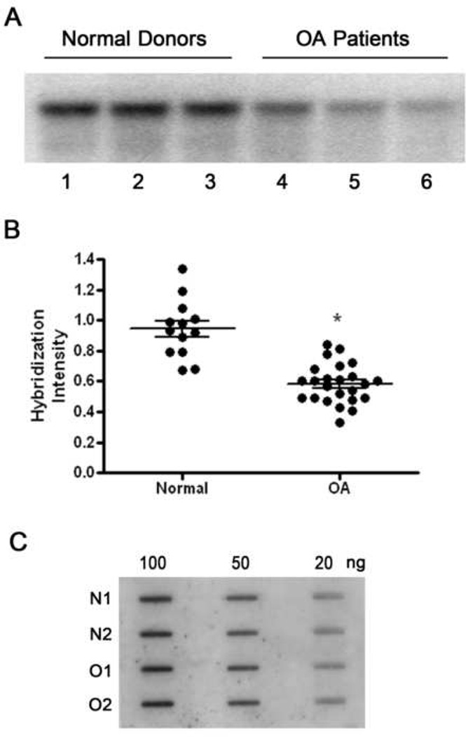
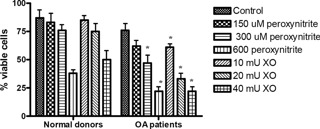
Methods: Human articular cartilage was isolated from knee joints of cadavers available through the Anatomical Gifts Program at the University of South Alabama (normal donors) or OA patients undergoing total knee replacement surgeries (OA patients). Total DNA was isolated from either chondrocytes released following collagenase digestion, or from first passage chondrocytes grown in culture and exposed to ROS or RNS. mtDNA integrity and repair capacity were analyzed by quantitative Southern blot analysis, using a mtDNA-specific radioactive probe. Cell viability was determined by the trypan blue exclusion method.

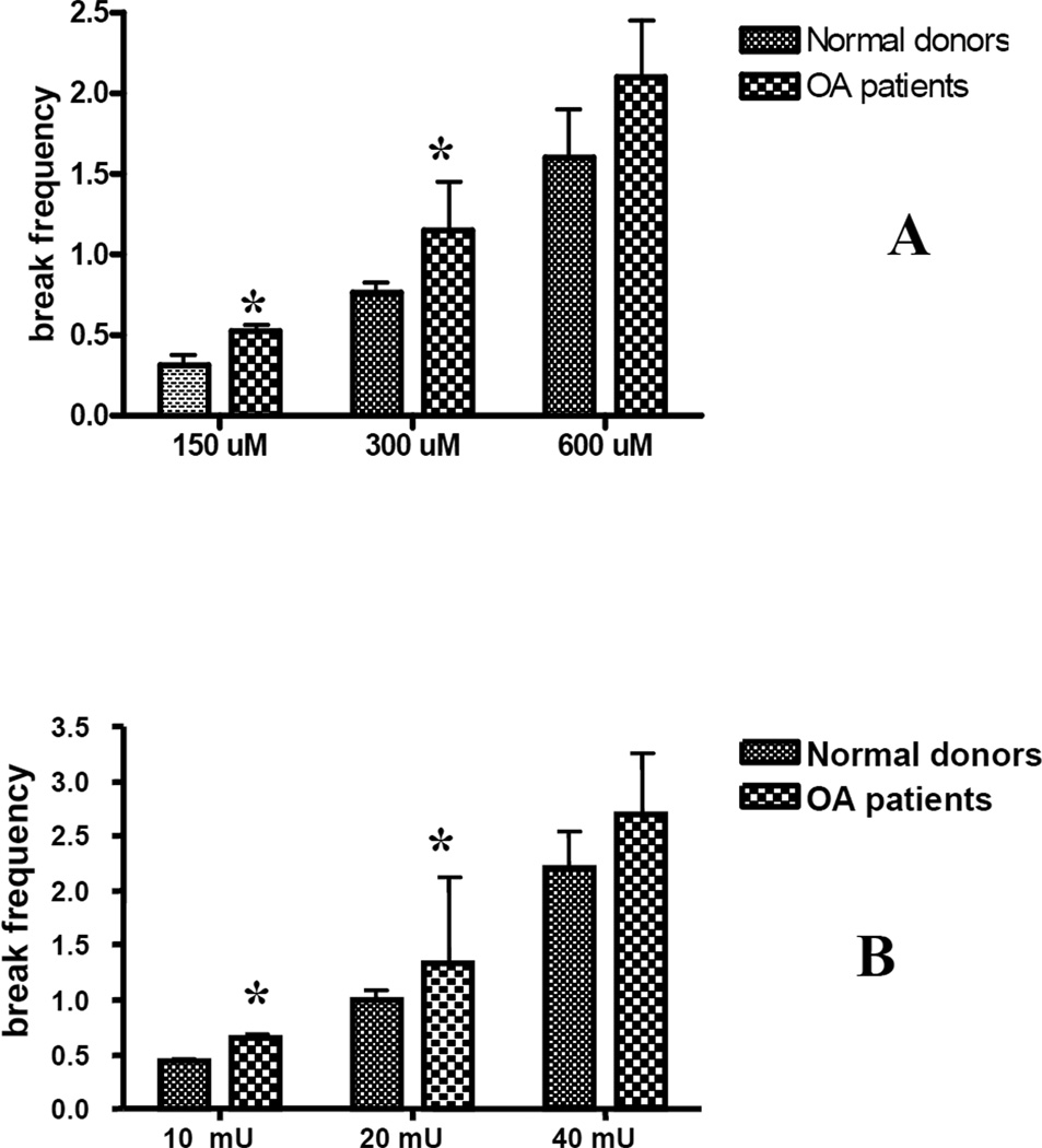
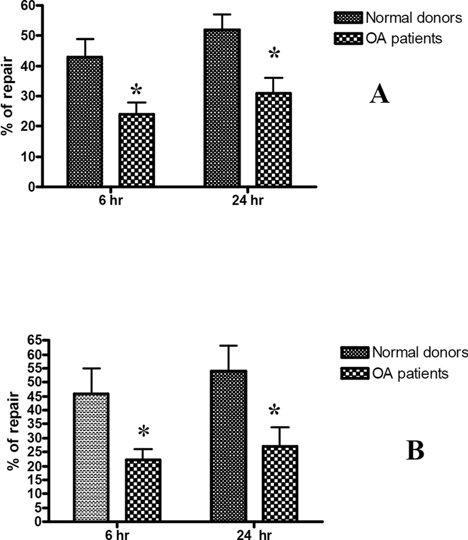
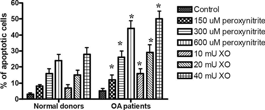
Results: mtDNA damage was found in chondrocytes from OA patients compared to normal donors. It was accompanied with reduced mtDNA repair capacity, cell viability, and increased apoptosis in OA chondrocytes following exposure to ROS and RNS.

Conclusions: These results indicate that mtDNA damage and poor mtDNA repair capacity for removing damage caused by oxidative stress may contribute to the pathogenesis of OA.


Figures

Fig. 1
Fig. 1. Mitochondrial DNA is damaged during the pathogenesis of OA
Fig. 1A is a representative autoradiogram from Southern blot analyses of mtDNA from three normal donors (lanes 1–3) and three OA patients (line 4–6). Human chondrocytes were lysed for DNA extraction following overnight digestion with collagenase B. The reduced intensity of the hybridization bands indicates that mtDNA damage has increased. Fig.1B displays a comparison of the hybridization band intensities obtained from the Southern blot analysis of 13 normal donors and 24 OA patients. * indicates a significant difference (P<0.05) between normal donors and OA patients. Fig.1C is a representative autoradiogram from slot-blot analyses performed on the DNA from normal donors and OA patients and shows that there is no variation in the amount of mtDNA between normal donors and OA patients, as well as between separate DNA samples.
Fig. 2
Fig. 2. Mitochondrial DNA in chondrocytes from OA patients is more sensitive to oxidative stress caused by exposure to ROS or RNS
Chondrocytes were exposed to increased concentrations of peroxynitrite (panel A) or xanthine oxidase with hypoxanthine (panel B) for 30 min. Cells were lysed, DNA was isolated and subjected to Southern blot analysis. An Increase in the break frequency indicates that more mtDNA damage has accumulated. The results were obtained from a minimum of 8 independent experiments, and the values represent the mean break frequency ± SEM. * indicates a significant difference (P<0.05) in damage in mtDNA in chondrocytes from OA patients compared to normal donors.
Fig. 3
Fig. 3. OA chondrocytes have a diminished capacity to repair oxidative damage
Primary human chondrocytes from OA patients and normal donors were exposed to 300 µM of peroxynitrite (panel A) or 20 mU xanthine oxidase /0.5 mM hypoxanthine for 30 min and allowed to repair for 6 and 24 h. Cells were lysed, DNA was isolated and subjected to Southern blot analysis. The results were obtained from a minimum of 7 independent experiments, and the values displayed represent the mean break frequency ± SEM. * indicates a significant difference (P<0.05) between OA chondrocytes and those isolated from normal donors.
Fig. 4
Fig. 4. Chondrocyte viability following exposure to ROS or RNS
Confluent cell cultures were exposed to increasing concentrations of xanthine oxidase or peroxynitrite for 30 min and left for recovery for 24 h. After 24h, cells were trypsinized away from the culture vessel, incubated with trypan blue and viewed by light microscopy to calculate the numbers of viable and dead cells. The results were obtained from a minimum of 6 independent experiments, and the values represent the mean percentage of viable cells ± SEM. * indicates a significant difference (P<0.05) in OA chondrocytes compared to those from normal donors. Note the diminished OA chondrocyte viability following exposure to both genotoxins.
Fig. 5
Fig. 5. The accumulation of apoptotic chondrocytes in cultures obtained from OA patients and normal donors following exposure to ROS or RNS
Twenty four hours following exposure to increasing concentrations of xanthine oxidase or peroxynitrite, cells were washed three times with PBS and fixed with 4% paraformaldehyde. The fixed cells were washed again with PBS and stained with 1 µg/ml DAPI. Stained cells were examined by fluorescent microscopy to identify apoptotic cells. To evaluate the percentage of apoptotic cells, 6–7 fields from each experimental condition were viewed in a blinded fashion until a total of 500 cells were observed. The results were obtained from a minimum of 6 independent experiments, and the values represent the mean percentage of apoptotic cells ± SEM. * indicates a significant difference (P<0.05) in the percentage of apoptotic cells in cultures from OA patients compared to normal donors. Note the increased in apoptosis in OA chondrocytes following exposure to both genotoxins.

References

    1. Henrotin YE, Bruckner P, Pujol JP. The role of reactive oxygen species in homeostasis and degradation of cartilage. Osteoarthritis Cartilage. 2003;11:747–755. - PubMed
    1. Mazzetti I, Grigolo B, Pulsatelli L, Dolzani P, Silvestri T, Roseti L, et al. Differential roles of nitric oxide and oxygen radicals in chondrocytes affected by osteoarthritis and rheumatoid arthritis. Clin Sci (Lond) 2001;101:593–599. - PubMed
    1. Fermor B, Weinberg JB, Pisetsky DS, Misukonis MA, Banes AJ, Guilak F. The effects of static and intermittent compression on nitric oxide production in articular cartilage explants. J Orthop Res. 2001;19:729–737. - PubMed
    1. Blanco FJ, Ochs RL, Schwarz H, Lotz M. Chondrocyte apoptosis induced by nitric oxide. Am J Pathol. 1995;146:75–85. - PMC - PubMed
    1. Farrell AJ, Blake DR, Palmer RM, Moncada S. Increased concentrations of nitrite in synovial fluid and serum samples suggest increased nitric oxide synthesis in rheumatic diseases. Ann Rheum Dis. 1992;51:1219–1222. - PMC - PubMed

Publication types