. 2008 Jul 1;105(26):9105-10.
doi: 10.1073/pnas.0800289105. Epub 2008 Jun 17.

Cyclic adenosine monophosphate differentiated beta-endorphin neurons promote immune function and prevent prostate cancer growth

Affiliations

Cyclic adenosine monophosphate differentiated beta-endorphin neurons promote immune function and prevent prostate cancer growth

Dipak K Sarkar et al. Proc Natl Acad Sci U S A. .

Abstract

Pituitary adenylate cyclase-activating peptide (PACAP), a cAMP-activating agent, is highly expressed in the hypothalamus during the period when many neuroendocrine cells become differentiated from the neural stem cells (NSCs). Activation of the cAMP system in rat hypothalamic NSCs differentiated these cells into beta-endorphin (BEP)-producing neurons in culture. When these in vitro differentiated neurons were transplanted into the paraventricular nucleus (PVN) of the hypothalamus of an adult rat, they integrated well with the surrounding cells and produced BEP and its precursor gene product, proopiomelanocortin (POMC). Animals with BEP cell transplants demonstrated remarkable protection against carcinogen induction of prostate cancer. Unlike carcinogen-treated animals with control cell transplants, rats with BEP cell transplants showed rare development of glandular hyperplasia, prostatic intraepithelial neoplasia (PIN), or well differentiated adenocarcinoma with invasion after N-methyl-N-nitrosourea (MNU) and testosterone treatments. Rats with the BEP neuron transplants showed increased natural killer (NK) cell cytolytic function in the spleens and peripheral blood mononuclear cells (PBMCs), elevated levels of antiinflammatory cytokine IFN-gamma, and decreased levels of inflammatory cytokine tumor necrosis factor-alpha (TNF-alpha) in plasma. These results identified a critical role for cAMP in the differentiation of BEP neurons and revealed a previously undescribed role of these neurons in combating the growth and progression of neoplastic conditions like prostate cancer, possibly by increasing the innate immune function and reducing the inflammatory milieu.


Conflict of interest statement

The authors declare no conflict of interest.

Figures

Fig. 1.
Fig. 1.
Immunocytochemical characterization of NSCs at different stages of differentiation. (A) Immunofluorescence staining of nestin, shown by arrows in NSCs at day 0. (B and C) At day 3, staining for vimentin (B) and α-internexin (C) in the early stage of differentiation. (D–F) At day 7, phase-contrast image (D) and staining for the neuronal marker NF-M (E) and BEP (F). (G) Control NSCs maintained in neuron culture medium without PACAP and cAMP showed no staining for BEP but showed DAPI staining. (H–L) At day 14, phase-contrast image (H), staining for neuronal markers MAP2 (I) and type III β-tubulin J, no staining for GFAP (K), and positive staining for BEP (L). Green staining is for NSCs or neural cell marker, red staining is for BEP, and blue staining is for nuclear DAPI. (Scale bars: 10 μm.)
Fig. 2.
Fig. 2.
Biochemical characterization of NSCs at different stages of differentiation. (A and B) BEP release (A) and POMC mRNA expression (B) in NSCs at different time periods of differentiation in the presence of PACAP (10 μM) and dbcAMP (10 μM). No BEP was detected in media samples collected from control cultures without vehicle (0-day) or the cultures treated with the differentiating agents for 3 days (3-day). (C and D) Dose-response and the synergistic effect of PACAP and dbcAMP on BEP neuron differentiation as determined by the ability of NSCs to release BEP (C) and express POMC mRNA (D) after treatment with the drug for 1 wk and then without the drugs for 1 wk. The control group was treated similarly with vehicle. (E and F) PgE1 (10 μM for 3 h)-induced BEP release (E) and POMC mRNA expression (F) in differentiated cells. Values are presented as a percentage of vehicle-treated control. n = 6–8. a, P < 0.05; b, P < 0.01; c, P < 0.001 vs. the rest.
Fig. 3.
Fig. 3.
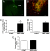
Determination of the viability of NSC-derived BEP cells transplanted in the PVN. Young male rats were transplanted with in vitro differentiated BEP cells or nonviable BEP cell (CONT) unilaterally in the PVN. The viability of these transplanted cells was determined by immunocytochemical (A and B) and biochemical (C–E) procedures. (A) Representative photographs showing BrdU-stained cells at the site of transplantation. (B) High-power view showing immunofluorescence staining for BrdU (green), BEP (red), and merged (yellow) of transplanted cells. Many BEP-stained cells show long processes (arrows). (Scale bars: 10 μm.) (C–E) POMC mRNA levels (C) and BEP levels in the PVN (D) and BEP levels in plasma (E) of animals with BEP cell or CONT transplants. n = 6–8. c, P < 0.001 vs. CONT.
Fig. 4.
Fig. 4.
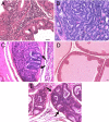
Evaluation of the effect of BEP cell transplants on the MNU and testosterone-induced prostate cancers. Adult male rats were transplanted with in vitro differentiated BEP cells or cortical cells (CONT) bilaterally in the PVN of male rats. These rats were then treated with MNU and testosterone treatments and used for determination of histopathology of prostates. (A–C) Prostates of rats transplanted with CONT showed lesions ranging from epithelial hyperplasia with mild atypia (A) to high-grade PIN (B) and occasionally invasive adenocarcinoma (shown by arrows; C). (D and E) Prostates of rats transplanted with BEP cells demonstrated very mild changes, ranging from minimal (D) to moderate hyperplasia (shown by arrows; E). (Scale bars: 10 μm.)
Fig. 5.
Fig. 5.
Evaluation of the effects of BEP cell transplants on immune functions. Rats were transplanted with BEP cells or cortical cells (CONT) into one PVN (+) or two PVN (++) for 4 wk. Shown are dose-response effects of BEP cell transplants on splenic NK cell cytolytic activity (A), PBMC-derived NK cell cytolytic activity (B), plasma IFN-γ (C), and plasma TNF-α (D). n = 5–7 rats. a, P < 0.05; b, P < 0.01; c, P < 0.001 vs. the CONT.

Similar articles

Cited by

References

    1. Settle M. The hypothalamus. Neonatal Netw. 2000;19:9–14. - PubMed
    1. van Eerdenburg FJ, Rakic P. Early neurogenesis in the anterior hypothalamus of the Rhesus monkey. Brain Res Dev Brain Res. 1994;79:290–296. - PubMed
    1. Altman J, Bayer SA. Development of the diencephalon in the rat. III. Ontogeny of the specialized ventricular linings of the hypothalamic third ventricle. J Comp Neurol. 1978;182:995–1015. - PubMed
    1. De A, Boyadjieva N, Pastorcic M, Reddy BV, Sarkar DK. cAMP and ethanol interact to control apoptosis and differentiation in hypothalamic β-endorphin neurons. J Biol Chem. 1994;269:26697–26705. - PubMed
    1. Plotsky PM, Thrivikraman KV, Meaney MJ. Central and feedback regulation of hypothalamic corticotropin releasing factor secretion. Ciba Found Symp. 1993;172:59–75. - PubMed

Publication types

MeSH terms