. 2008 Jun 24;105(25):8691-6.
doi: 10.1073/pnas.0803732105. Epub 2008 Jun 18.

Ablation of thymic export causes accelerated decay of naive CD4 T cells in the periphery because of activation by environmental antigen

Affiliations

Ablation of thymic export causes accelerated decay of naive CD4 T cells in the periphery because of activation by environmental antigen

Christine Bourgeois et al. Proc Natl Acad Sci U S A. .

Abstract

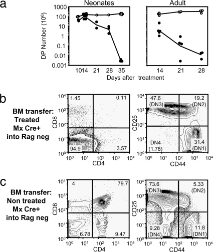
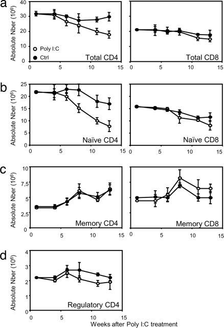
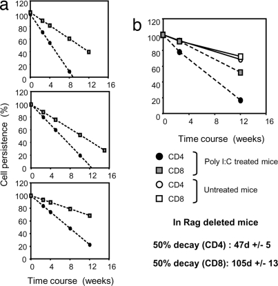
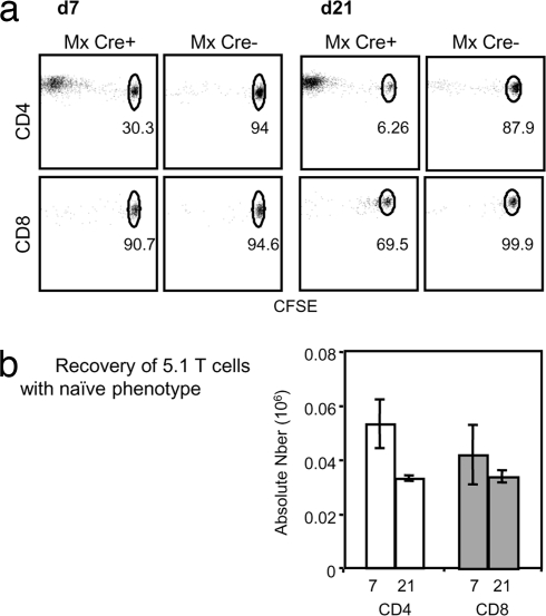
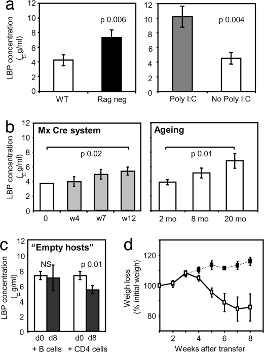
A model of chemical thymectomy by inducible Rag ablation was used to study peripheral T cell homeostasis. Induction of Rag ablation was efficient and complete, leading to cessation of thymic T cell production within 3-4 weeks. The decay of peripheral T cells became apparent with a delay of an additional 2-3 weeks and was entirely accounted for by loss of naïve T cells, whereas numbers of memory phenotype and regulatory T cells were not decreased. Naïve CD4 T cells decayed with an average half-life of 50 days, whereas naïve CD8 T cells exhibited a considerably longer half-life. The rapid decay of naïve CD4 T cells was not caused by intrinsic survival differences compared with naïve CD8 T cells, but was caused by changes in the lymphopenic environment resulting in higher microbial load and consequential activation. This finding suggests that in lymphopenic conditions involving compromised thymic function replenishment and survival of a naïve CD4 T cell repertoire may be severely curtailed because of chronic activation. Such a scenario might play a role in the aging immune system and chronic viral infection, such as HIV infection, and contribute to loss of CD4 T cells and impaired immune function. As our data show, continued replenishment with cells from the thymus seems to be required to maintain efficient gut mucosal defense.


Conflict of interest statement

The authors declare no conflict of interest.

Figures

Fig. 1.
Fig. 1.
Efficiency of the deletion. (a) Absolute cell numbers of DP thymocytes after type I IFN treatment of 3-day-old MxCre- Rag2 fl/− (open symbols) or MxCre+ Rag2 fl/− (filled symbols) (Left) or after poly(I:C) treatment of 8- to 10-week-old mice (Right). (b and c) Dot plots of gated CD45.2 cells from thymus of bone marrow chimeras MxCre+ Rag2fl/−-treated with poly(I:C) 4 weeks before bone marrow isolation (b) or MxCre+ Rag2fl/− untreated bone marrow (c) injected into 5 Gy irradiated Rag2-deficient B6 CD45.1 hosts. (Left) Dot plots show CD8 vs. CD4 profiles. (Right) Dot plots show CD25 vs. CD44 profiles of the DN population with stages DN1 to DN4 marked.
Fig. 2.
Fig. 2.
Peripheral T cell survival in the absence of thymic export. Peripheral T cell numbers were determined in poly(I:C)-treated (○) or untreated (●) MxCre+ Rag2fl/− mice. Numbers of CD4 (Left) and CD8 (Right) peripheral T cells (a), naïve T cells (b), memory T cells (c), and regulatory CD4 T cells (d) are shown. Mean values and SDs for three mice per time point are shown. These data are representative of three experiments using three mice per group for each experiment.
Fig. 3.
Fig. 3.
Decay of naïve CD4 and CD8 T cells after thymus ablation. CD4 and CD8 T cell decay was extrapolated from the numbers of naïve CD4 and CD8 T cells recovered over a course of 15 weeks after poly(I:C) treatment and plotted against values obtained from untreated Mx Cre+ Rag2fl/− littermates, as shown in Fig. 2b. Cell numbers obtained 5 weeks after treatment were set as 100% value on the y axis and 0 on the x axis. (a) Trend lines for CD4 (circles) and CD8 (squares) T cell decay in poly(I:C)-treated Mx Cre+ Rag2fl/− mice in three independent experiments. (b) Average trend lines for CD4 (circles) and CD8 (squares) T cell decay in poly(I:C)-treated (filled symbols) or untreated (open symbols) Mx Cre+ Rag2fl/− mice. Note that naïve CD4 and CD8 T cells from untreated mice exhibit identical decay and therefore show overlapping trend lines on the graph. Error bars were omitted from the summary graph in the interest of visual clarity.
Fig. 4.
Fig. 4.
CD4 T cells, but not CD8 T cells, proliferate in poly(I:C)-treated MxCre+ hosts. (a) Proliferation and recovery of sorted B6 CD45.1 naïve CD4 T cells (Upper) or naïve CD8 T cells (Lower) after transfer into poly(I:C)-treated Mx Cre+ Rag2fl/− or Mx Cre Rag2fl/− control mice. CFSE profiles were obtained 7 and 21 days after transfer. (b) Absolute numbers of recovered donor CD4 (open bars) or CD8 T cells (filled bars) with a naïve phenotype 7 and 21 days after transfer into poly(I:C)-treated Mx Cre+ Rag2fl mice. This experiment is representative of three experiments using three mice per group and per time point.
Fig. 5.
Fig. 5.
LBP levels in serum. (a) LBP concentration in the sera of 8-week-old B6 mice (open bar) and Rag2-deficient mice (black bar) (Left) and LBP concentration in serum of poly(I:C)-treated mice (gray bar) or untreated Mx Cre+ Rag2fl/− mice (open bar) 15–20 weeks after treatment (Right). This experiment is representative of two experiments using three to five mice per group. (b) LBP concentration in sera of poly(I:C)-treated MxCre+ Rag2fl/− mice at different time points after poly(I:C) treatment (Left) and LBP concentration in sera of B6 mice at 2, 8, and 20 months of age (Right). (c) LPB concentration in sera of Rag-deficient mice injected with purified B cells or naïve CD4 T cells (5 × 10⁶ per mouse) before cell transfer at day 0 (open bars) or 8 days after cell transfer (filled bars). (d) Transfer of 1.5 × 10⁶ naïve CD4 T cells isolated from poly(I:C)-treated (■) or untreated (□) Mx Cre+ Rag2fl/− mice into Rag-deficient hosts. The graph shows percentage of weigh loss in the adoptive hosts. These data are representative of two experiments using at least four mice per group.

Similar articles

Cited by

References

    1. Tanchot C, Rocha B. The peripheral T cell repertoire: Independent homeostatic regulation of virgin and activated CD8+ T cell pools. Eur J Immunol. 1995;25:2127–2136. - PubMed
    1. Jameson SC. Maintaining the norm: T cell homeostasis. Nat Rev Immunol. 2002;2:547–556. - PubMed
    1. Mackall CL, Gress RE. Thymic aging and T cell regeneration. Immunol Rev. 1997;160:91–102. - PubMed
    1. Ge Q, Hu H, Eisen HN, Chen J. Different contributions of thymopoiesis and homeostasis-driven proliferation to the reconstitution of naive and memory T cell compartments. Proc Natl Acad Sci USA. 2002;99:2989–2994. - PMC - PubMed
    1. Berzins SP, Boyd RL, Miller JF. The role of the thymus and recent thymic migrants in the maintenance of the adult peripheral lymphocyte pool. J Exp Med. 1998;187:1839–1848. - PMC - PubMed

Publication types

LinkOut - more resources