. 2008 Jun 24;105(25):8760-5.
doi: 10.1073/pnas.0803545105. Epub 2008 Jun 18.

Endocannabinoid signaling controls pyramidal cell specification and long-range axon patterning

Affiliations

Endocannabinoid signaling controls pyramidal cell specification and long-range axon patterning

Jan Mulder et al. Proc Natl Acad Sci U S A. .

Abstract

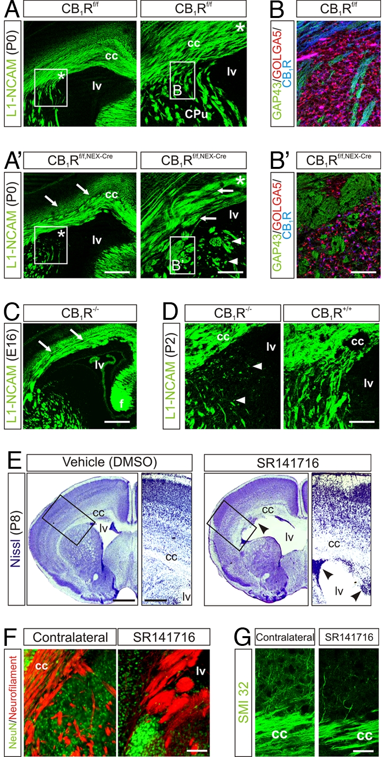
Endocannabinoids (eCBs) have recently been identified as axon guidance cues shaping the connectivity of local GABAergic interneurons in the developing cerebrum. However, eCB functions during pyramidal cell specification and establishment of long-range axonal connections are unknown. Here, we show that eCB signaling is operational in subcortical proliferative zones from embryonic day 12 in the mouse telencephalon and controls the proliferation of pyramidal cell progenitors and radial migration of immature pyramidal cells. When layer patterning is accomplished, developing pyramidal cells rely on eCB signaling to initiate the elongation and fasciculation of their long-range axons. Accordingly, CB(1) cannabinoid receptor (CB(1)R) null and pyramidal cell-specific conditional mutant (CB(1)R(f/f,NEX-Cre)) mice develop deficits in neuronal progenitor proliferation and axon fasciculation. Likewise, axonal pathfinding becomes impaired after in utero pharmacological blockade of CB(1)Rs. Overall, eCBs are fundamental developmental cues controlling pyramidal cell development during corticogenesis.


Conflict of interest statement

The authors declare no conflict of interest.

Figures

Fig. 1.
Fig. 1.
CB1R localization in the developing brain. (A–G) In situ hybridization demonstrating the spatial and temporal distribution of CB1R mRNA in the mouse brain. Arrows in C and E and G denote CB1R mRNA hybridization signal in pyramidal cells and SVZ progenitors, respectively. (H and I) Distribution of CB1R mRNA in human fetal brain. Nissl/AChE histochemistry reveals territorial boundaries. (J–M) DAGLβ and CB1R expression in mouse VZ/SVZ. Cortical Tbr2+ projection neurons migrating toward the CP express CB1Rs. (N–R) CB1Rs are selectively enriched in axons of cortical projection neurons. Arrows indicate corticothalamic axons, and arrowheads identify axons committed to the fibria. (Scale bars: E–G and K–M, 30 μm; N–R, 85 μm; A–D, H, and I, 100 μm.) See SI Text for abbreviations.
Fig. 2.
Fig. 2.
eCBs regulate neural progenitor proliferation in cortical VZ/SVZ. (A) CB1R deletion significantly reduces the rate of neural progenitor proliferation, as defined by the density of BrdU+ cells in the VZ/SVZ. (B) Conversely, elevated eCB levels in FAAH−/− mice (14) significantly increase neural progenitor proliferation. (C) Conditional CB1R deletion in subcortical SVZ progenitors (arrows) (16) decreases the rate of Ki67+ progenitor proliferation (n = 3 per genotype). **, P < 0.01, compared with wild-type littermates. (Scale bar, 100 μm.)
Fig. 3.
Fig. 3.
eCBs control radial migration of pyramidal cell progenitors. (A) Cortical distribution of neurons at P2.5 whose progenitors were BrdU labeled at E14.5. Note the migration arrest of a population of neural progenitors in deep cortical layers of CB1R−/− mice. (B) Cortical progenitor distribution in FAAH−/− mice was assessed as above. Cell counts were performed in grouped cortical layers defined as equal binned areas. (C) Distribution of GFP+ cells in brain slices from E14.5 mouse embryos after ex vivo electroporation of VZ progenitors with pCIG2-GFP. Slices were maintained for 48 h in the presence of HU-210 (1 μM) or URB597 (1 μM) in vitro. Cumulative cell counts were obtained in CP, IZ, and VZ/SVZs. **, P < 0.01; *, P < 0.05, compared with wild-type littermates or control treatment. (see Tables S1 and S2 for statistical analysis). (Scale bars: A and B, 75 μm; C, 35 μm.)
Fig. 4.
Fig. 4.
eCB signaling controls pyramidal cell morphogenesis. (A and B) Quantitative morphometry of axon development of VGLUT1+ pyramidal cells after 3-day treatment in vitro. Bracketed numbers indicate population sizes (see also Table S3). (C–E) DAGLα and β undergo axonal targeting and exhibit differential distribution (C). Whereas DAGLα concentrates in axonal varicosities, DAGLβ is distributed uniformly along axons (arrows in D). (F) In axonal growth cones, DAGLα is targeted to filopodia (arrows), whereas DAGLβ concentrates in the axon stem with a clear demarcation from the growth cones (arrowheads). (G) DAGL inhibition by O-3841 significantly reduces VGLUT1 expression in pyramidal cells by 6 days in vitro. O-3841 effects were prevented by exogenous application of AEA. *, P < 0.05, compared with control. (Scale bars: A, 25 μm; C, 15 μm; D–F, 3 μm.)
Fig. 5.
Fig. 5.
Genetic and pharmacological ablation of CB1Rs leads to axon fasciculation deficits. (A) Conditional deletion of CB1Rs in pyramidal cells evokes fasciculation deficits in L1-NCAM+ long-range axons. Arrows point to enlarged fascicles. Asterisk in open boxes denotes the general localization of insets showing the lack of fiber invasion in dorsal striatum. (B and B′) Aberrant axon bundles in dorsal striatum. (C) Axonal deficits in CB1R−/− brains at E16 are reminiscent of those seen in neonatal conditional mutants. (D) Impaired axonal targeting toward the striatum persists in CB1R−/− until P2. (E) In utero SR141716 infusion induces ventricle enlargement, migration arrest, and cortical delamination. Arrowheads indicate clusters of progenitor cells in the ventricular zone. Open boxes denote the position of insets. (F) Neurofilament M staining reveals fasciculation deficits in the subventricular corpus callosum after SR141716 infusion. (G) SR141716 decreases the commitment of pyramidal cell axons to descending projections. (Scale bars: A, C, and E, 250 μm; A Inset and D, 100 μm; B′ and G, 30 μm; F, 50 μm.)

References

    1. Rakic P. A century of progress in corticoneurogenesis: From silver impregnation to genetic engineering. Cereb Cortex. 2006;16(Suppl 1):i3–i17. - PubMed
    1. Fishell G, Hanashima C. Pyramidal neurons grow up and change their mind. Neuron. 2008;57:333–338. - PubMed
    1. Lopez-Bendito G, et al. Tangential neuronal migration controls axon guidance: a role for neuregulin-1 in thalamocortical axon navigation. Cell. 2006;125:127–142. - PMC - PubMed
    1. Domenici MR, et al. Cannabinoid receptor type 1 located on presynaptic terminals of principal neurons in the forebrain controls glutamatergic synaptic transmission. J Neurosci. 2006;26:5794–5799. - PMC - PubMed
    1. Kawamura Y, et al. The CB1 cannabinoid receptor is the major cannabinoid receptor at excitatory presynaptic sites in the hippocampus and cerebellum. J Neurosci. 2006;26:2991–3001. - PMC - PubMed

Publication types

Substances

LinkOut - more resources