. 2008 Aug 29;283(35):24194-201.
doi: 10.1074/jbc.M802869200. Epub 2008 Jun 18.

Resveratrol inhibits cardiac hypertrophy via AMP-activated protein kinase and Akt

Affiliations

Resveratrol inhibits cardiac hypertrophy via AMP-activated protein kinase and Akt

Anita Y M Chan et al. J Biol Chem. .

Abstract

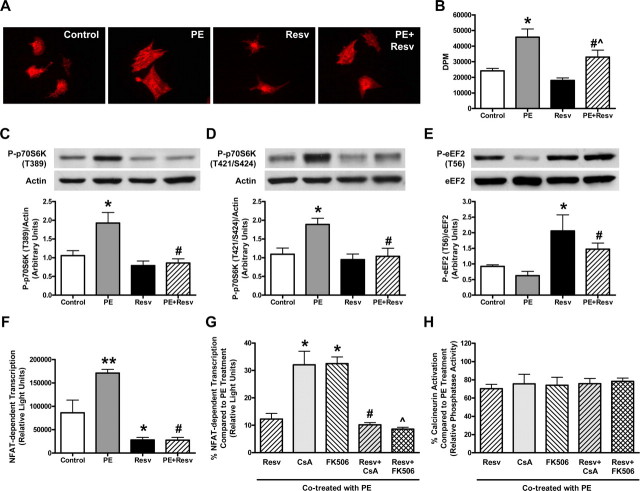
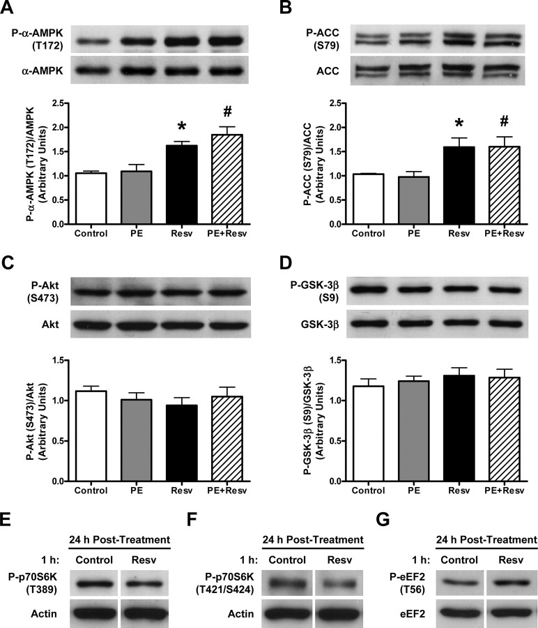
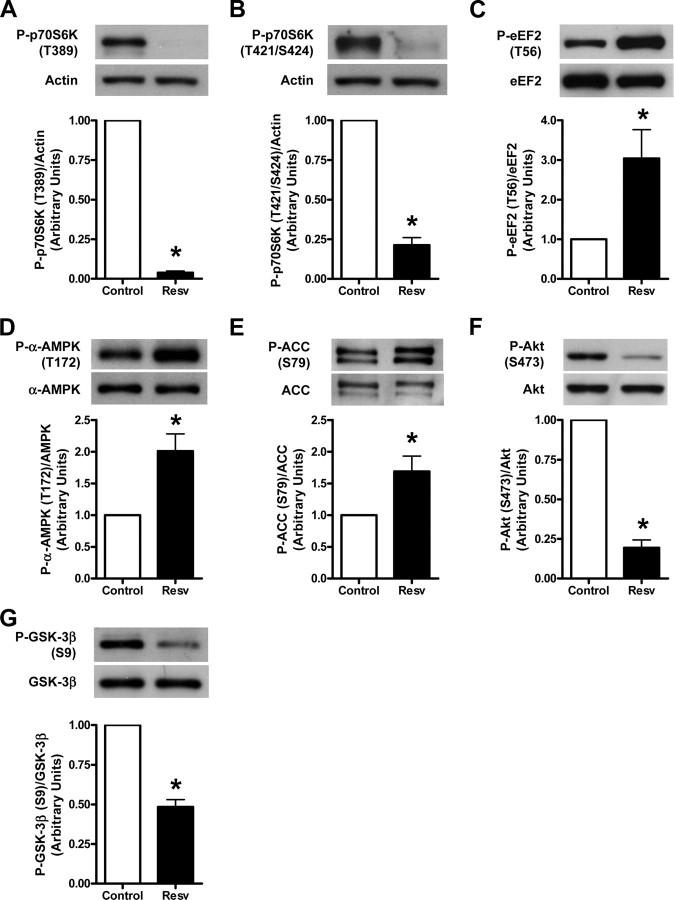
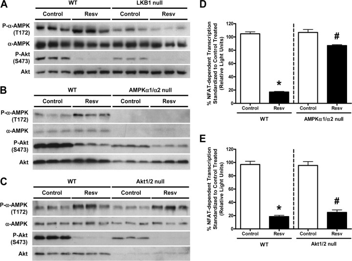
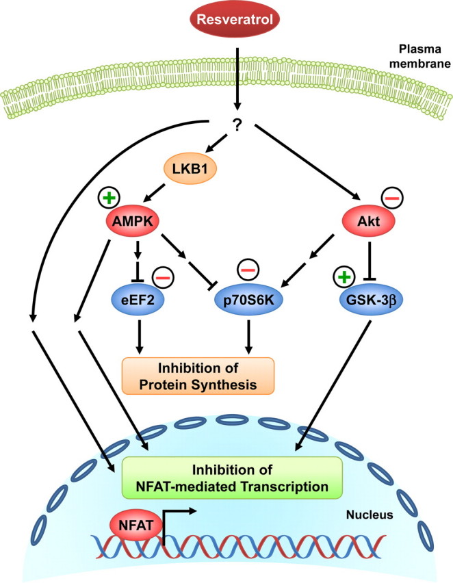
Whereas studies involving animal models of cardiovascular disease demonstrated that resveratrol is able to inhibit hypertrophic growth, the mechanisms involved have not been elucidated. Because studies in cells other than cardiomyocytes revealed that AMP-activated protein kinase (AMPK) and Akt are affected by resveratrol, we hypothesized that resveratrol prevents cardiac myocyte hypertrophy via these two kinase systems. Herein, we demonstrate that resveratrol reduces phenylephrine-induced protein synthesis and cell growth in rat cardiac myocytes via alterations of intracellular pathways involved in controlling protein synthesis (p70S6 kinase and eukaryotic elongation factor-2). Additionally, we demonstrate that resveratrol negatively regulates the calcineurin-nuclear factor of activated T cells pathway thus modifying a critical component of the transcriptional mechanism involved in pathological cardiac hypertrophy. Our data also indicate that these effects of resveratrol are mediated via AMPK activation and Akt inhibition, and in the case of AMPK, is dependent on the presence of the AMPK kinase, LKB1. Taken together, our data suggest that resveratrol exerts anti-hypertrophic effects by activating AMPK via LKB1 and inhibiting Akt, thus suppressing protein synthesis and gene transcription.


Figures

FIGURE 1.
FIGURE 1.
Resveratrol blunts phenylephrine-induced cardiac myocyte hypertrophy and protein synthesis via the p70S6K, eEF2, and calcineurin-NFAT signaling pathways. A, qualitative representation of the cell size of neonatal rat cardiac myocytes treated with ethanol (Control) or 50 μm resveratrol (Resv), in the absence or presence of 10 μm phenylephrine (PE) for 24 h. Changes in cell size were visualized following fixation on coverslips using mouse anti-α-actinin and Texas Red-conjugated donkey anti-mouse antibodies. B, measurement of protein synthesis using [3H]phenylalanine incorporation with counts expressed in DPM (disintegrations per minute). Values are means ± S.E. with each experiment performed in duplicate (n = 4). *, p < 0.01 versus Control; #, p < 0.05 versus PE; ^, p < 0.05 versus Resv; assessed by ANOVA and Bonferroni multiple comparisons test. C-E, representative immunoblot and densitometry of cellular extracts from cardiac myocytes treated as described above. Values are expressed as means ± S.E. and analyzed with the Kruskal-Wallis test followed by Dunn's multiple comparisons test. C, cell lysates blotted with anti-phospho-p70S6K (T389) and anti-actin antibodies; *, p < 0.05 versus Control; #, p < 0.01 versus PE (n = 9-10). D, cell lysates blotted with anti-phospho-p70S6K (T421/S424) and anti-actin antibodies; *, p < 0.05 versus Control; #, p < 0.05 versus PE (n = 5-6). E, cell lysates blotted with anti-phospho-eEF2 (T56) and anti-eEF2 antibodies; *, p < 0.05 versus Control; #, p < 0.01 versus PE (n = 8-9). F, cellular extracts of cardiac myocytes infected with Ad.NFAT-Luc-Promoter adenovirus and treated as described above were assessed for NFAT-dependent transcription, measured as luciferase activity expressed in relative light units. Values are means ± S.E. with each sample assessed in duplicate (n = 5); **, p < 0.001 versus Control; *, p < 0.05 versus Control; #, p < 0.001 versus PE, analyzed by ANOVA and Bonferroni multiple comparisons test. G and H, myocytes infected with Ad.NFAT-Luc-Promoter adenovirus were treated with 500 ng/ml (416 nm) of CsA or 150 ng/ml (182 nm) of 506 with or without 50 μm Resv, in the presence of 10 μm PE for 24 h. G, NFAT-dependent transcription measured from cellular extracts is presented as a percentage (%) compared with PE treatment, where values are means ± S.E. with each sample assessed in duplicate (n = 4); *, p < 0.001 versus Resv; #, p < 0.001 versus CsA; ^, p < 0.001 versus FK506, analyzed by ANOVA and Bonferroni multiple comparisons test. H, calcineurin activity measured from cellular extracts is presented as a percentage (%) compared with PE treatment, where values are means ± S.E. with each sample assessed in duplicate (n = 4-5), analyzed by ANOVA.
FIGURE 2.
FIGURE 2.
Chronic resveratrol treatment increases AMPK and ACC phosphorylation without changing Akt and GSK-3β phosphorylation, while acute treatment has lasting effects on p70S6K and eEF2 phosphorylation levels. A-D, representative immunoblot and densitometry of cellular extracts from neonatal rat cardiac myocytes treated with ethanol (Control) or 50 μm resveratrol (Resv), in the absence or presence of 10 μm phenylephrine (PE) for 24 h. Values are expressed as means ± S.E. and analyzed with the Kruskal-Wallis test followed by Dunn's multiple comparisons test. A, cell lysates blotted with anti-phospho-α-AMPK (T172) and anti-α-AMPK antibodies; *, p < 0.01 versus Control; #, p < 0.05 versus PE (n = 9-10). B, cell lysates blotted with anti-phospho-ACC (S79) antibody and peroxidase-labeled streptavidin, which detects both isoforms of ACC (upper and lower bands) and were quantified together; *, p < 0.05 versus Control; #, p < 0.05 versus PE (n = 5-7). C, cell lysates blotted with anti-phospho-Akt (S473) and anti-Akt antibodies (n = 9). D, cell lysates blotted with anti-phospho-GSK-3β (S9) and anti-GSK-3β antibodies (n = 9). E-G, representative immunoblot of cellular extracts from neonatal rat cardiac myocytes treated with ethanol (Control) or 100 μm Resv for 1 h, followed by 24 h of incubation with serum-free medium (n = 4). E, cell lysates blotted with anti-phospho-p70S6K (T389) and anti-actin antibodies, (F) anti-phospho-p70S6K (T421/S424) and anti-actin antibodies, and (G) anti-phospho-eEF2 (T56) and anti-eEF2 antibodies.
FIGURE 3.
FIGURE 3.
Acute treatment with resveratrol has dramatic effects on the p70S6K and eEF2 signaling pathways, while affecting both AMPK and Akt signaling pathways. Representative immunoblot and densitometry of cellular extracts from neonatal rat cardiac myocytes treated with ethanol (Control) or 100 μm resveratrol (Resv) for 1 h. Values are expressed as means ± S.E. and analyzed with Student's one sample t test as control was set to 1. A, cell lysates blotted with anti-phospho-p70S6K (T389) and anti-actin antibodies, *, p < 0.0001 versus Control (n = 4); (B) anti-phospho-p70S6K (T421/S424) and anti-actin antibodies, *, p < 0.001 versus Control (n = 4); (C) anti-phospho-eEF2 (T56) and anti-eEF2 antibodies, *, p < 0.05 versus Control (n = 9); (D) anti-phospho-α-AMPK (T172) and anti-α-AMPK antibodies, *, p < 0.05 versus Control (n = 6); (E) antiphospho-ACC (S79) antibody and peroxidase-labeled streptavidin, with both ACC bands being quantified together, *, p < 0.05 versus Control (n = 7); (F) anti-phospho-Akt (S473) and anti-Akt antibodies, *, p < 0.001 versus Control (n = 4); and (G) anti-phospho-GSK-3β (S9) and anti-GSK-3β antibodies, *, p < 0.01 versus Control (n = 4).
FIGURE 4.
FIGURE 4.
The effect of resveratrol on AMPK phosphorylation is mediated by LKB1 without cross-talk between AMPK and Akt, and its effect on NFAT-dependent transcription is mainly dependent on AMPK not Akt. A-C, representative immunoblots of cellular extracts from mouse embryonic fibroblasts (MEFs) treated with ethanol (Control) or 100 μm resveratrol (Resv) for 1 h, blotted with anti-phospho-α-AMPK (T172), anti-α-AMPK, anti-phospho-Akt (S473), and anti-Akt antibodies. A, wild-type (WT) and LKB1-null MEFs; B, WT and AMPKα1/α2-null MEFs; and C, WT and Akt1/2-null MEFs were utilized (n = 3). D and E, AMPKα1/α2-null and Akt1/2-null MEFs with their respective WT MEFs were infected with Ad.NFAT-Luc-Promoter adenovirus and then treated with ethanol (Control) or 100 μm Resv for 1 h. NFAT-dependent transcription measured from cellular extracts is presented as a percentage (%) compared with control-treated, where values are means ± S.E. D,*, p < 0.01 versus WT control-treated; #, p < 0.05 versus AMPKα1/α2-null control-treated (n = 4-6), assessed by two-tailed Mann-Whitney test. E,*, p < 0.001 versus WT control-treated; #, p < 0.001 versus Akt1/2-null control-treated (n = 3), assessed by Student's unpaired two-tailed t test.
FIGURE 5.
FIGURE 5.
Proposed mechanisms of the anti-hypertrophic effects of resveratrol. Resveratrol, either directly or indirectly, activates AMPK via LKB1 and inhibits Akt. This results in a reduction of p70S6K and eEF2 activities and the subsequent inhibition of protein synthesis. The reduced Akt activity relieves the suppression of GSK-3β activity, which may also contribute to the inhibition of NFAT-mediated transcription. AMPK activation by resveratrol appears to mediate a major component of the diminished NFAT transcriptional activity in the nucleus via inhibition of calcineurin and yet to be identified mechanism(s). Resveratrol treatment may also decrease NFAT-mediated transcription via pathways independently of AMPK and Akt. Together, all of these resveratrol-mediated effects contribute to the inhibition of cardiac myocyte hypertrophy.

Similar articles

Cited by

References

    1. Benjamin, E. J., and Levy, D. (1999) Am. J. Med. Sci. 317 168-175 - PubMed
    1. Wilkins, B. J., Dai, Y. S., Bueno, O. F., Parsons, S. A., Xu, J., Plank, D. M., Jones, F., Kimball, T. R., and Molkentin, J. D. (2004) Circ. Res. 94 110-118 - PubMed
    1. Wilkins, B. J., and Molkentin, J. D. (2004) Biochem. Biophys. Res. Commun. 322 1178-1191 - PubMed
    1. Horman, S., Beauloye, C., Vertommen, D., Vanoverschelde, J. L., Hue, L., and Rider, M. H. (2003) J. Biol. Chem. 278 41970-41976 - PubMed
    1. Chan, A. Y., Soltys, C. L., Young, M. E., Proud, C. G., and Dyck, J. R. (2004) J. Biol. Chem. 279 32771-32779 - PubMed

Publication types

MeSH terms

LinkOut - more resources