. 2008 Jul;132(1):13-28.
doi: 10.1085/jgp.200709913. Epub 2008 Jun 18.

Direct regulation of BK channels by phosphatidylinositol 4,5-bisphosphate as a novel signaling pathway

Affiliations

Direct regulation of BK channels by phosphatidylinositol 4,5-bisphosphate as a novel signaling pathway

Thirumalini Vaithianathan et al. J Gen Physiol. 2008 Jul.

Abstract

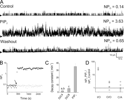
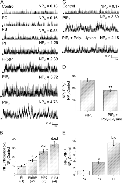
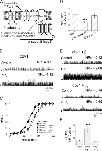
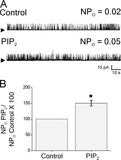
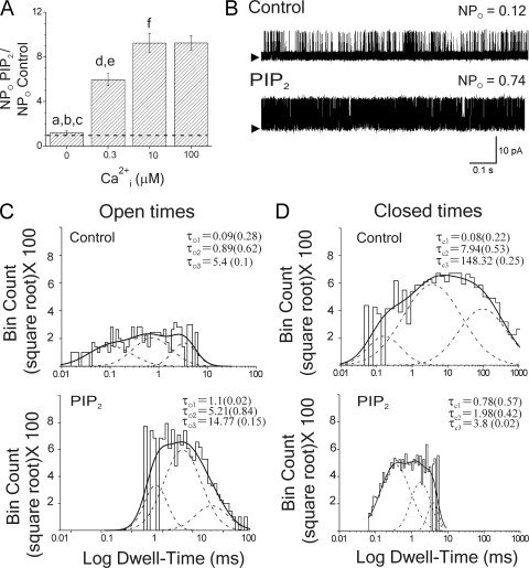
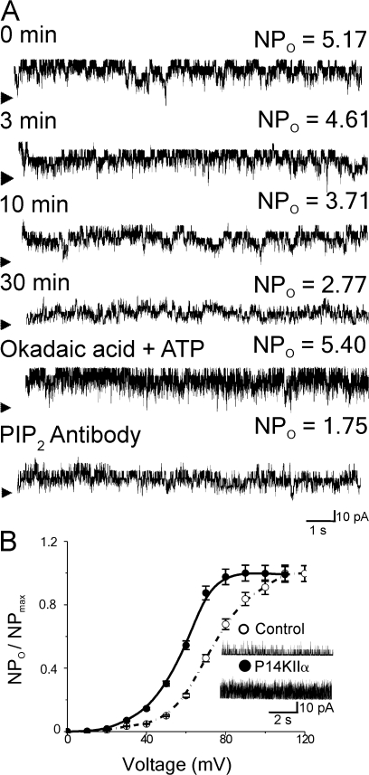
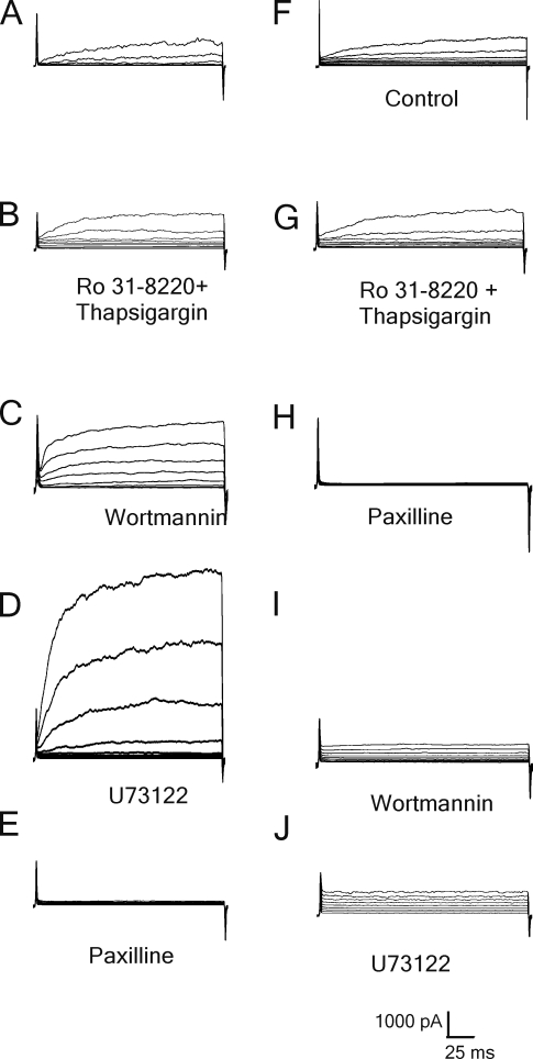
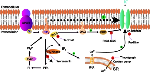
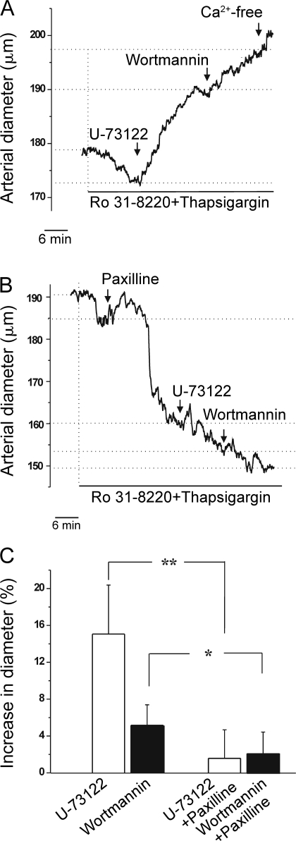
Large conductance, calcium- and voltage-gated potassium (BK) channels are ubiquitous and critical for neuronal function, immunity, and smooth muscle contractility. BK channels are thought to be regulated by phosphatidylinositol 4,5-bisphosphate (PIP(2)) only through phospholipase C (PLC)-generated PIP(2) metabolites that target Ca(2+) stores and protein kinase C and, eventually, the BK channel. Here, we report that PIP(2) activates BK channels independently of PIP(2) metabolites. PIP(2) enhances Ca(2+)-driven gating and alters both open and closed channel distributions without affecting voltage gating and unitary conductance. Recovery from activation was strongly dependent on PIP(2) acyl chain length, with channels exposed to water-soluble diC4 and diC8 showing much faster recovery than those exposed to PIP(2) (diC16). The PIP(2)-channel interaction requires negative charge and the inositol moiety in the phospholipid headgroup, and the sequence RKK in the S6-S7 cytosolic linker of the BK channel-forming (cbv1) subunit. PIP(2)-induced activation is drastically potentiated by accessory beta(1) (but not beta(4)) channel subunits. Moreover, PIP(2) robustly activates BK channels in vascular myocytes, where beta(1) subunits are abundantly expressed, but not in skeletal myocytes, where these subunits are barely detectable. These data demonstrate that the final PIP(2) effect is determined by channel accessory subunits, and such mechanism is subunit specific. In HEK293 cells, cotransfection of cbv1+beta(1) and PI4-kinaseIIalpha robustly activates BK channels, suggesting a role for endogenous PIP(2) in modulating channel activity. Indeed, in membrane patches excised from vascular myocytes, BK channel activity runs down and Mg-ATP recovers it, this recovery being abolished by PIP(2) antibodies applied to the cytosolic membrane surface. Moreover, in intact arterial myocytes under physiological conditions, PLC inhibition on top of blockade of downstream signaling leads to drastic BK channel activation. Finally, pharmacological treatment that raises PIP(2) levels and activates BK channels dilates de-endothelized arteries that regulate cerebral blood flow. These data indicate that endogenous PIP(2) directly activates vascular myocyte BK channels to control vascular tone.


Figures

Figure 1.
Figure 1.
PIP2 readily activates myocyte BK channels when accessing the channel from the inner membrane leaflet. (A) Unitary currents obtained before (top), after a 5-min bath application of 10 μM PIP2 (diC16) (middle), and after washout for >30 min (bottom) of I/O patches. Arrowheads, baseline; upward deflections, channel openings; n = 5. (B) Steady-state activity (NPo) time course from two I/O patches. An arrow highlights the time at which one of the patches (•) was switched from control to PIP2-containing solution. The other patch was continuously exposed to control (○). (C) Kinetics of reversibility of diC4, diC8, and PIP2 action. Decay constant values were obtained by single exponential fittings of NPo vs. time plots. (a) diC4 vs. diC8: P < 0.001; (b) diC4 vs. diC8: P < 0.001; (c) diC8 vs. PIP2: P < 0.001, n = 3–4. In A and B, V = 40 mV, Ca2+i= 0.3 μM; n = 3. (D) NPo responses to 10 μM PIP2 from I/O, O/O, and C/A patches. Each point = one patch/myocyte. Dotted line, control; V = 10–60 mV; Ca2+i = 0.3 μM; n = 4–6.
Figure 2.
Figure 2.
Negative charge and inositol moiety both contribute to phosphoinositide activation of myocyte BK channels. (A) Unitary currents from I/O patches exposed to control or phospholipids that differ in headgroup structure. Arrowheads, baseline; upward deflections, channel openings. (B) BK channel potentiation is a direct function (r = 0.99) of negative charge in the phospholipid headgroup. (a) PI vs. PI5P: P < 0.001; (b) PI vs. PIP2: P < 0.001; (c) PI5P vs. PIP2: P < 0.001; (d) PI vs. PIP3: P < 0.001; (e) PI5P vs. PIP3: P < 0.001; (f) PIP2 vs. PIP3: P < 0.05; n = 3–12. (C) I/O recordings and (D) averaged data show that coapplication of 0.1 mg/ml poly-l-lysine reduces PIP2 activation of BK channels; n = 3. (E) PS and PI (net charge ≈ −1) cause more robust activation than that evoked by the neutral PC. However, PI is more effective than PS. (a) PC vs. PS: P < 0.05; (b) PC vs. PI: P < 0.001; (c) PS vs. PI: P < 0.001; n = 3–6. All phospholipid species had dipalmitoyl chains. V = 40 mV; Ca2+i = 0.3 μM.
Figure 3.
Figure 3.
Cbv1 is sufficient to support PIP2 action, which is amplified by β1 (but not β4) subunits. (A) BK channel dimer made of channel-forming (cbv1) and auxiliary β(1–4) subunits. The RKK to AAA mutation in the cbv1 S6–S7 linker is shown in bold. (B) Unitary currents from an I/O patch expressing cbv1 in the absence (top) and presence (bottom) of PIP2. Arrowheads, baseline; upward deflections, channel openings. (C) Averaged G-voltage macroscopic current data fitted to Boltzmann functions from wt cbv1, RKKcbv1AAA, and K239cbv1A in the absence and presence of PIP2; the lipid causes a parallel leftward shift in wt and K239cbv1A but not in the RKKcbv1AAA mutant; n = 4–6. (D) PIP2-induced increase in NPo is significantly reduced in the RKKcbv1AAA mutant when compared with wt cbv1 or the K239cbv1A mutant; ***, P < 0.001; n = 4. (E). Unitary currents from I/O patches coexpressing cbv1+β1 (top) or cbv1+β4 subunits (bottom) in the absence and presence of PIP2. Arrowheads, baseline; upward deflections, channel openings. (F) Averaged PIP2 responses of cbv1, cbv1+β1, and cbv1+β4; n = 4–6. For B and E, arrows, baseline. For A, B, and D–F, V = 40 mV; Ca2+i = 0.3 μM.
Figure 4.
Figure 4.
PIP2 action on native BK channels in isolated skeletal muscle myocytes. (A) Unitary currents from an I/O patch obtained before (top) and after (bottom) a 5-min bath application of 10 μM PIP2 show that the phosphoinositide causes an increase in BK NPo that is markedly reduced when compared with that evoked by PIP2 in vascular myocyte BK channels (Fig. 1, A and C). Arrowheads, baseline; upward deflections, channel openings. (B) Averaged PIP2 responses of native skeletal muscle BK channels; *, P < 0.05; n = 4; V = 40 mV, Ca2+i = 10 μM.
Figure 5.
Figure 5.
PIP2 activates the BK channel by amplifying Ca2+-driven gating, resulting in modifications of both open and closed times. (A) PIP2 activation of cbv1 is negligible at zero nominal Ca2+i and reaches a maximum at 10 μM Ca2+i; (a) zero Ca2+i vs. 0.3 μM Ca2+i: P < 0.001; (b) zero Ca2+i vs. 10 μM Ca2+i: P < 0.001; (c) zero Ca2+i vs. 100 μM Ca2+i: P < 0.001; (d) 0.3 μM Ca2+i vs. 10 μM Ca2+i: P < 0.001; (e) 0.3 μM Ca2+i vs. 100 μM Ca2+i: P < 0.001; n = 3–12. (B) Single channel records in the absence (top) and presence (bottom) of 10 μM PIP2 show that the lipid increases Po from 0.06 to 0.43 (616% of control). Records were low-passed at 7 kHz and digitized at 35 kHz. Upward deflections: channel openings. (C) Open and (D) closed time distributions in control (top) and PIP2 (bottom) from records shown in B. Each life time constant (τ) is shown in milliseconds, with its contribution to the total fit in parentheses. Each component of a fit is shown with a dotted line, and the composite fit is shown with a solid line; 40 mV; Ca2+i = 0.3 μM.
Figure 6.
Figure 6.
Endogenous PIP2 maintains native BK channel activity. (A) Time course of vascular myocyte BK NPo from an I/O patch. Intervals indicate time after patch excision. Arrowheads, baseline; upward deflections, channel openings. Bath application of 0.5 mM ATP and 0.1 μM okadaic acid (OA) increases NPo ×5.4 times. Subsequent application of PIP2 monoclonal antibodies (1:1,000) drastically reduces NPo. V = 40 mV; Ca2+i = 3 μM. (B) Cotransfection of HEK cells with PI4KαII and cbv1+β1 channel subunits causes a parallel leftward shift in the activation–voltage plot when compared with currents mediated by cbv1+β1 alone; n = 5–7.
Figure 7.
Figure 7.
Endogenous PIP2 activates native BK currents in the presence of blockers of PLC-mediated PIP2 downstream products. Perforated patch recordings from two (A–E and F–J) freshly isolated cerebral artery myocytes bathed in physiological saline solution (PSS; composition in Materials and methods). Total outward K+ currents were recorded in the continuous presence of 5 mM 4-AP and 0.1 mM niflumic acid. Bath application of 2 μM Ro31-8220 and 0.2 μM thapsigargin increases mean outward current by 95% (B vs. A, and G vs. F). Subsequent inhibition of PI3 kinase by 5 nM wortmannin further increases current by 184% from control (C). Inhibition of PLC by 25 μM U73122 drastically increases current (D), likely due to buildup of PIP2 in the membrane. The current is blocked by 0.3 μM paxilline (E and H), indicating it is mediated by BK channels. Preapplication of paxilline, a selective BK channel blocker, prevents both wortmannin (I) and U73122 (J) actions (n = 5–6).
Figure 8.
Figure 8.
Indirect (classical) and direct (novel) mechanisms of PIP2 action on BK currents. Activated PLC via Gq, determined by either ligand binding to a Gq-coupled receptor (GPCR) or constitutive activity, cleaves PIP2 into PIP3, IP3, and DAG. IP3 releases Ca2+ from the sarcoplasmic reticulum (SR), raising Ca2+i, whereas DAG activates PKC in the presence of Ca2+. BK channel modulation by PKC and Ca2+i controls the degree of vascular myocyte contraction. Therefore, by generating DAG and IP3, PIP2 indirectly modulates BK channels and, thus, vascular tone. We demonstrate that PIP2 itself and other membrane phosphoinositides directly activate BK channels, that is, in the absence of cell integrity or organelles or in the continuous presence of cytosolic messengers. This activation involves the negative charges of the phosphoinositide headgroup and the sequence of positive residues RKK in the cbv1 S6–S7 cytosolic linker. However, PIP2 activation is drastically and distinctly amplified by the channel accessory subunit of the β1 type, which is abundantly expressed in smooth muscle. In intact myocytes, inhibition of PLC by U73122 increases paxilline-sensitive BK currents in the presence of a PI3 kinase blocker (wortmannin), PKC inhibitor (Ro 31-8220), and a selective blocker of the sarcoplasmic reticulum Ca2+-ATPase (thapsigargin). Under these conditions, PLC inhibition increases membrane PIP2 (Narayanan et al., 1994), which leads to increased BK current in the intact cell (see main text).
Figure 9.
Figure 9.
Endogenous PIP2 dilates cerebral arteries via BK channels. Diameter traces from de-endothelized arteries after developing myogenic tone. (A) In the presence of 2 μM Ro 31-8220 and 0.2 μM thapsigargin, PLC inhibition (25 μM U73122) causes a robust dilation (+15.1%), which is further increased by 5 nM wortmannin. Maximal dilation is evoked by Ca2+-free solution. (B) BK channel block (0.3 μM paxilline) reduces diameter (−13%) and almost totally blunts U73122 and wortmannin actions. In A and B, a vertical line indicates the time at which Ro 31-8220 and thapsigargin were applied; horizontal lines help to visualize diameter changes. (C) Averaged diameters; **, P < 0.01; *, P < 0.05; n = 4.

Comment in

  • PIP2 PIP2 hooray for maxi K+.
    Rittenhouse AR. Rittenhouse AR. J Gen Physiol. 2008 Jul;132(1):5-8. doi: 10.1085/jgp.200810053. Epub 2008 Jun 18. J Gen Physiol. 2008. PMID: 18562503 Free PMC article. No abstract available.

Similar articles

Cited by

References

    1. Arcaro, A., and M.P. Wymann. 1993. Wortmannin is a potent phosphatidylinositol 3-kinase inhibitor: the role of phosphatidylinositol 3,4,5-trisphosphate in neutrophil responses. Biochem. J. 296:297–301. - PMC - PubMed
    1. Bao, L., and D.H. Cox. 2005. Gating and ionic currents reveal how the BKCa channel's Ca2+ sensitivity is enhanced by its β1 subunit. J. Gen. Physiol. 126:393–412. - PMC - PubMed
    1. Barman, S., S. Zhu, and R. White. 2004. PKC activates BKCa channels in rat pulmonary arterial smooth muscle via cGMP-dependent protein kinase. Am. J. Physiol. Lung Cell Mol. Physiol. 286:L149–L155. - PubMed
    1. Behrens, R., A. Nolting, F. Reimann, M. Schwarz, R. Waldschutz, and O. Pongs. 2000. hKCNMB3 and hKCNMB4, cloning and characterization of two members of the large-conductance calcium-activated potassium channel β subunit family. FEBS Lett. 474:99–106. - PubMed
    1. Brauchi, S., G. Orta, C. Mascayano, M. Salazar, N. Raddatz, H. Urbina, E. Rosenmann, F. Gonzalez-Nilo, and R. Latorre. 2007. Dissection of the components for PIP2 activation and thermosensation in TRP channels. Proc. Natl. Acad. Sci. USA. 104:10246–10251. - PMC - PubMed

Publication types

MeSH terms

Substances