Comparative Study
. 2008 Jun 18;28(25):6372-82.
doi: 10.1523/JNEUROSCI.1054-08.2008.

Preferential reactivation of motivationally relevant information in the ventral striatum

Affiliations
Comparative Study

Preferential reactivation of motivationally relevant information in the ventral striatum

Carien S Lansink et al. J Neurosci. .

Abstract

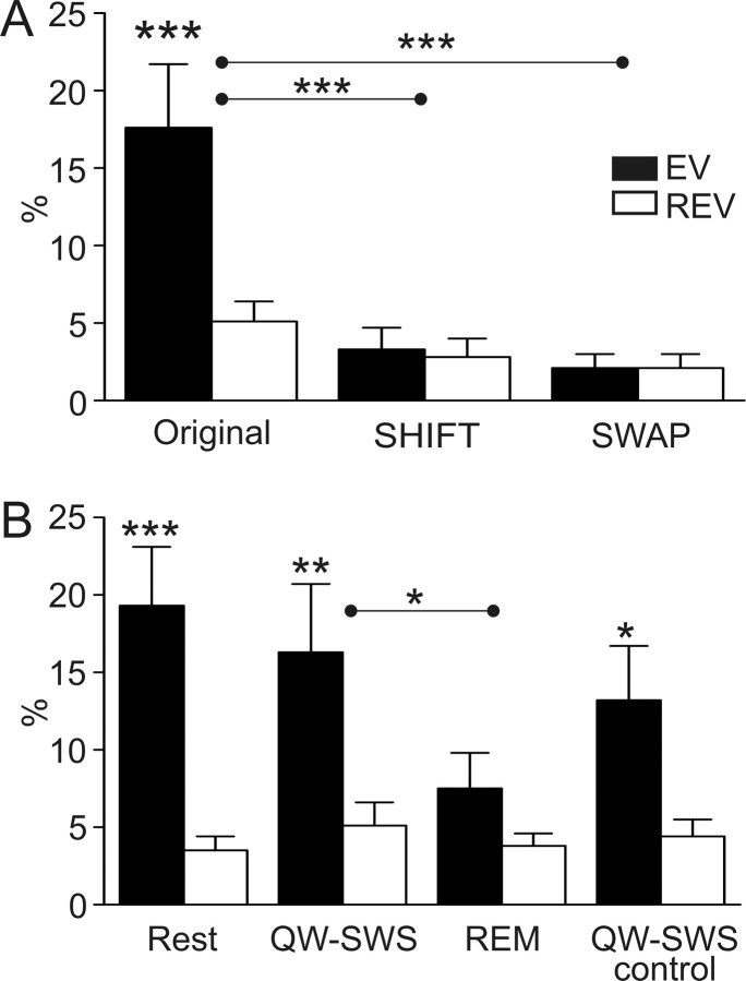
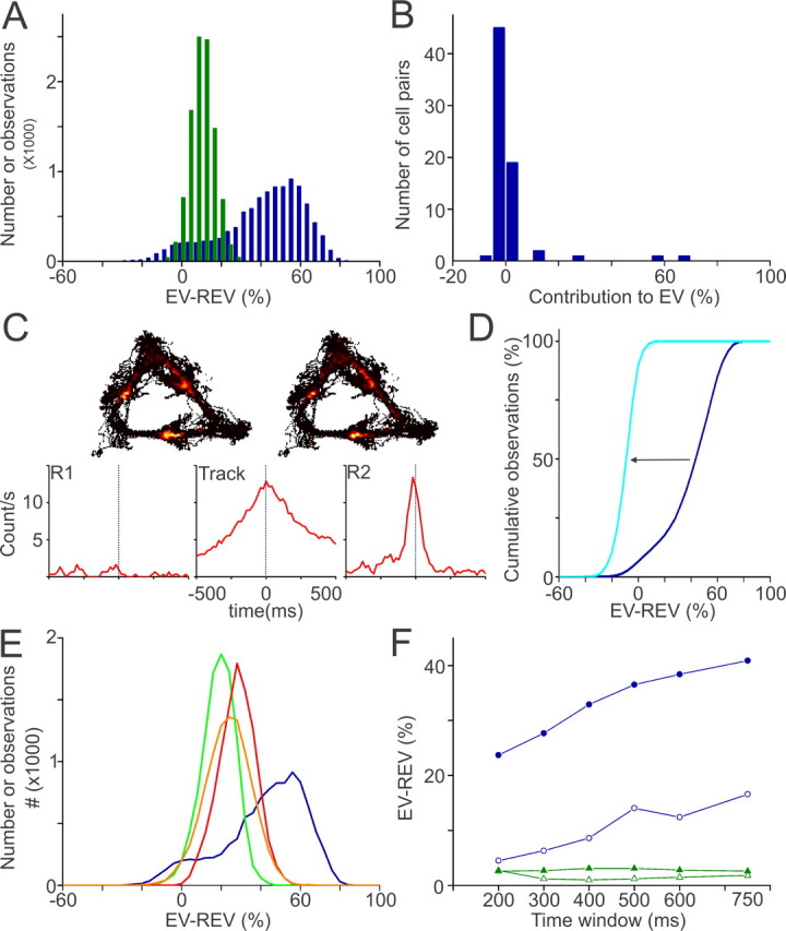
Spontaneous "off-line" reactivation of neuronal activity patterns may contribute to the consolidation of memory traces. The ventral striatum exhibits reactivation and has been implicated in the processing of motivational information. It is unknown, however, whether reactivating neuronal ensembles specifically recapitulate information relating to rewards that were encountered during wakefulness. We demonstrate a prolonged reactivation in rat ventral striatum during quiet wakefulness and slow-wave but not rapid eye movement sleep. Reactivation of reward-related information processed in this structure was particularly prominent, and this was primarily attributable to spike trains temporally linked to reward sites. It was accounted for by small, strongly correlated subgroups in recorded cell assemblies and can thus be characterized as a sparse phenomenon. Our results indicate that reactivated memory traces may not only comprise feature- and context-specific information but also contain a value component.


Figures

Figure 1.
Figure 1.
A, Example of a tetrode recording: projection plots of cluster isolation, waveforms, and interspike interval (ISI) histograms. Spikes belonging to separate clusters were identified according to multiple waveform features including area under the curve (plotted in this figure), peak amplitude, and principal components. X, Y, and Z correspond to three of four leads of the tetrode. The color of the waveforms shown in the first and third column (bottom) corresponds to the colors of the individual clusters in the projection plots. Top, The black diagonal band corresponds to unclassified events, including noise. Bottom, In the second and fourth column, ISI histograms are shown for each unit represented to the left, with ISI counts on the ordinate and interval duration on the abscissa. B, Schematic representation of the endpoints of the tetrode tips. The tetrode endings were in the ventral striatum approximately between 2.2 and 1.2 anterior to bregma and between 1.6 and 3.0 laterally compared with an atlas of the rat brain (Paxinos and Watson, 1996).
Figure 2.
Figure 2.
Ventral striatal units showing firing patterns associated with one or more reward sites. Rats ran along a triangular track to obtain three types of reward (S, sucrose; V, vanilla; C, chocolate). Top panels, Spatial distribution of firing rates of two individual neurons. A, B, Local firing rates ranged from 0 to maxima of 19 Hz (A) and 18 Hz (B). Bottom panels, Perievent time histograms for both cells synchronized on reward site arrivals. Rows represent different reward types, whereas columns differentiate between the presence and absence of reward. Firing rate is in bins of 250 ms. The red ticks indicate arrivals at other reward sites. A, Neuron showing an increased firing rate shortly before and after arriving at the sucrose reward site, mainly when a reward was obtained. B, Neuron increased its firing rate before arrival at two sites differentially for the rewarded versus nonrewarded condition. The response at the vanilla site was stronger than at the chocolate site. The rat may have detected the availability of reward before arriving at the well by visual or olfactory cues, although reward cups were filled outside the rat's field of view.
Figure 3.
Figure 3.
A–C, Examples of the firing patterns of a ventral striatal ensemble, plotted in parallel with the local field potential recorded near the hippocampal fissure (A) and the pyramidal cell layer (B, C). A, As the rat is running on the track, theta oscillations were observed in the hippocampal field potentials. The bottom and top traces represented raw and filtered (6–10 Hz) field potentials, respectively. In a period of 15 s, the rat crossed four reward sites (s, sucrose; v, vanilla dessert; c, chocolate mousse), two of which were baited (indicated with green arrows) and two of which were not (red arrows). Each row in the plot below the field potential traces represents a single unit; its spikes are marked by specifically colored dots. Note the high variability in the firing patterns around each reward site arrival but also the positively correlated firing of the “green” (4) and “blue” (5) units. B, The hippocampal local field potential during QW-SWS is dominated by large irregular activity interleaved with sharp wave–ripple complexes. The bottom and top traces represent raw and filtered (100–250 Hz) field potentials. Note the concurrent firing of the green (4) and blue (5) units (two high contributors) indicated by black arrows. C, Enlargement of a segment of the LFP traces and spike patterns shown in B. The firing of units 4 and 5 as well as spikes of unit 8 are aligned to identified ripples in the hippocampal LFP, which are marked with asterisks.
Figure 4.
Figure 4.
Reactivation in the ventral striatum: general characteristics and sleep phases. A, The EV exceeded the REV when compared across sessions (original: ***p < 0.001; n = 30). Reactivation disappeared when the temporal alignment between spike trains was disrupted (SHIFT, EVshift vs EVoriginal, ***p < 0.001) or when cell identities were randomized (SWAP, EVswap vs EVoriginal, ***p < 0.001). B, Reactivation observed during QW-SWS sleep (**p < 0.02) was not different from the reactivation found for the entire rest episodes, which included all episodes of motionless behavior (rest, n = 16 sessions). In contrast, REM sleep periods did not show reactivation and EV values were significantly lower than for QW-SWS (*p < 0.05). This lack of reactivation cannot be ascribed to the relatively late occurrence of REM sleep periods after sleep onset because post-REM QW-SWS periods showed significant reactivation (QW-SWS control, *p < 0.05). The strength of the observed reactivation during these intervals was not significantly different from the complete QW-SWS periods. Error bars indicate SEM.
Figure 5.
Figure 5.
Strong reactivation of ventral striatal units showing reward-related firing patterns. A, Bootstrap distributions of EV − REV values of reward-related correlates (RRU; blue) and non-reward-related correlates (NRU; green). The RRU distribution was broad and bimodal, indicating inhomogeneous contributions of cell pairs to reactivation. B, Distribution of the contributions of individual RRU cell pairs to their session EV. Only a small fraction of the RRU cell pairs contributed >10% (5 of 70; high contributors). C, Rate maps of two neurons constituting a highly contributing pair. Cross-correlograms show that firing patterns of these cells, recorded on different tetrodes, were highly correlated in time on the track and in postbehavioral but not in prebehavioral rest. D, Reactivation disappeared after exclusion of highly contributing pairs from the total RRU group, as shown by a leftward shift in the cumulative EV − REV distribution (blue → light blue). E, Mean reactivation in the RRU group was a factor of 1.5–3.5 higher than in control groups matched for recording session (green), firing rate (orange), and strength of Pearson's correlation coefficients (red). F, EV − REV values plotted as function of the width of the time window before and after reward site visits or intervals. Spike patterns of RRUs (blue) that occurred in close temporal association to reward site visits (filled circles) were reactivated more strongly than those that occurred in intervals (open circles). Reactivation was not observed for the NRU group across the same time windows (green).

Comment in

References

    1. Battaglia FP, Sutherland GR, McNaughton BL. Hippocampal sharp wave bursts coincide with neocortical “up-state” transitions. Learn Mem. 2004;11:697–704. - PMC - PubMed
    1. Buzsáki G. Hippocampal sharp waves: their origin and significance. Brain Res. 1986;398:242–252. - PubMed
    1. Corbit LH, Muir JL, Balleine BW. The role of the nucleus accumbens in instrumental conditioning: evidence of a functional dissociation between accumbens core and shell. J Neurosci. 2001;21:3251–3260. - PMC - PubMed
    1. Dahan L, Astier B, Vautrelle N, Urbain N, Kocsis B, Chouvet G. Prominent burst firing of dopaminergic neurons in the ventral tegmental area during paradoxical sleep. Neuropsychopharmacology. 2007;32:1232–1241. - PubMed
    1. Dalley JW, Lääne K, Theobald DE, Armstrong HC, Corlett PR, Chudasama Y, Robbins TW. Time-limited modulation of appetitive Pavlovian memory by D1 and NMDA receptors in the nucleus accumbens. Proc Natl Acad Sci USA. 2005;102:6189–6194. - PMC - PubMed

Publication types

LinkOut - more resources