Comparative Study
. 2008 Jun 18;28(25):6402-6.
doi: 10.1523/JNEUROSCI.1204-08.2008.

GluR6/KA2 kainate receptors mediate slow-deactivating currents

Affiliations
Comparative Study

GluR6/KA2 kainate receptors mediate slow-deactivating currents

Andrea Barberis et al. J Neurosci. .

Abstract

Kainate receptors (KARs) are ionotropic glutamate receptors contributing to EPSCs with a slow-decaying component that is likely essential for synaptic integration. The slow kinetics of KAR-EPSCs markedly contrasts with the fast kinetics reported for recombinant KARs expressed in heterologous systems, for reasons that remain unexplained. Here we have studied the properties of recombinant heteromeric GluR6/KA2 receptors, which compose synaptic KARs. We report that, in response to brief glutamate applications, currents mediated by recombinant GluR6/KA2 receptors, but not GluR6 receptors, decay with a time course similar to KAR-EPSCs. Model simulations suggest that, after brief agonist exposures, GluR6/KA2 currents undergo slow deactivation caused by the stabilization of partially bound open states. We propose, therefore, that the GluR6/KA2 gating features could contribute to the slow KAR-EPSC decay kinetics.


Figures
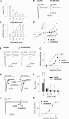
Figure 1.
Figure 1.
Properties of GluR6- and GluR6/KA2-mediated currents evoked by long glutamate pulses. A, Dependence of the current peak amplitude on GluR6:KA2 cDNA ratio. Currents were elicited by glutamate pulses (1 mm, 100 ms). B, Typical GluR6 and GluR6/KA2 current responses to glutamate (1 mm, 100 ms). GluR6 and KA2 subunits were transfected at the ratio 1:3. C, Dependence of the rectification index (ratio between the current obtained at membrane potential +120 mV and −120 mV) on GluR6:KA2 cDNA ratio. D, Current–voltage (I–V) curves for current mediated by GluR6 (black triangles) and GluR6/KA2 (gray triangles). GluR6 and KA2 cDNAs were transfected at the ratio 1:3. E, F, Typical GluR6-mediated (E) and GluR6/KA2-mediated (F) currents elicited by glutamate (1 mm and 10 μm). G, Dose–response curve for GluR6 (black triangles) and GluR6/KA2 receptors (gray triangles). H, Same traces as in E and F normalized and superimposed. I, Summary of the desensitization kinetics at different [Glu]. *p < 0.05. J, Typical example of GluR6 and GluR6/KA2 current responses to paired-pulse protocol (1 mm, 100 ms pulse length, 1 s gap). K, Time course of the recovery from desensitization (GluR6 and GluR6/KA2 receptors, black and gray triangles, respectively). In paired-pulse experiments, we only compared traces showing the same peak amplitude when evoked by either 1.5 or 100 ms glutamate pulses. τdes, desensitization time constant; fract., fractional.
Figure 2.
Figure 2.
GluR6/KA2 decay kinetics depends on stimulation pulse length. A, B, GluR6 currents and GluR6/KA2 currents evoked by either long (100 ms) or brief (1.5 ms) glutamate pulses. C, Paired pulses for GluR6- and GluR6/KA2-mediated currents (1 mm, 1.5 ms pulse length, 300 ms gap). D, Summary of the decay kinetics for GluR6 and GluR6/KA2 currents (black and gray bars, respectively) evoked by increasing glutamate pulse duration. E, Time course of recovery from desensitization for GluR6 and GluR6/KA2 receptors (black and gray squares, respectively) obtained with 1.5 ms pulses superimposed on that obtained with 100 ms pulses (same as that shown in Fig. 1M; black and gray triangles for GluR6 and GluR6/KA2 receptors, respectively). fract., Fractional.
Figure 3.
Figure 3.
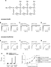
Model simulation. A, Kinetic scheme previously proposed by Robert and Howe (2003). The rate constants used to simulate the behavior of GluR6 and GluR6/KA2 receptors are listed in supplemental Table 1 (available at www.jneurosci.org as supplemental material). Simulated current traces were obtained by activating 1000 channels at the holding potential of −90 mV. When brief (1.5 ms) pulses were applied, the input waveform was trapezoidal, matching the whole-cell depolarization time course induced by 1.5 ms application of 25 mm potassium concentration saline, as detailed in supplemental Figure 1 (available at www.jneurosci.org as supplemental material). B, E, Simulated GluR6- and GluR6/KA2-mediated current evoked by 1 mm, 100 ms glutamate pulse (left) and by 1 mm, 1.5 ms, glutamate pulse (right). C, F, Simulated paired-pulse protocol for GluR6 and GluR6/KA2 receptors [100 ms, 1 mm, 300 ms gap (left) and 1.5 ms, 1 mm, 300 ms gap (right)]. D, G, Simulated GluR6- and GluR6/KA2-mediated current evoked by a synaptic-like glutamate exposure. The synaptic-like glutamate pulse was assumed to be exponential (see supplemental Model Simulations, available at www.jneurosci.org as supplemental material). H, Summary of the decay time constants for simulated GluR6 and GluR6/KA2 currents elicited by brief (1.5 ms), long (100 ms), and synaptic-like glutamate pulses. I, Time course of the recovery from desensitization for GluR6 and GluR6/KA2 receptors obtained with 1.5 ms pulses and 100 ms pulses (same color and symbol code as in Fig. 2E). fract., Fractional.

Similar articles

Cited by

References

    1. Bowie D, Garcia EP, Marshall J, Traynelis SF, Lange GD. Allosteric regulation and spatial distribution of kainate receptors bound to ancillary proteins. J Physiol (Lond) 2003;547:373–385. - PMC - PubMed
    1. Bureau I, Dieudonne S, Coussen F, Mulle C. Kainate receptor-mediated synaptic currents in cerebellar Golgi cells are not shaped by diffusion of glutamate. Proc Natl Acad Sci USA. 2000;97:6838–6843. - PMC - PubMed
    1. Castillo PE, Malenka RC, Nicoll RA. Kainate receptors mediate a slow postsynaptic current in hippocampal CA3 neurons. Nature. 1997;388:182–186. - PubMed
    1. Cho CH, St-Gelais F, Zhang W, Tomita S, Howe JR. Two families of TARP isoforms that have distinct effects on the kinetic properties of AMPA receptors and synaptic currents. Neuron. 2007;55:890–904. - PubMed
    1. Clements JD, Lester RA, Tong G, Jahr CE, Westbrook GL. The time course of glutamate in the synaptic cleft. Science. 1992;258:1498–1501. - PubMed

Publication types

Substances

LinkOut - more resources