Comparative Study
. 2008 Jun 18;28(25):6439-52.
doi: 10.1523/JNEUROSCI.0700-08.2008.

A dlx2- and pax6-dependent transcriptional code for periglomerular neuron specification in the adult olfactory bulb

Affiliations
Comparative Study

A dlx2- and pax6-dependent transcriptional code for periglomerular neuron specification in the adult olfactory bulb

Monika S Brill et al. J Neurosci. .

Abstract

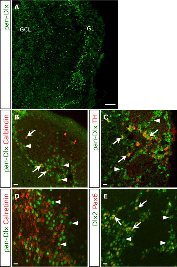
Distinct olfactory bulb (OB) interneurons are thought to become specified depending on from which of the different subregions lining the lateral ventricle wall they originate, but the role of region-specific transcription factors (TFs) in the generation of OB interneurons diversity is still poorly understood. Despite the crucial roles of the Dlx family of TFs for patterning and neurogenesis in the ventral telencephalon during embryonic development, their role in adult neurogenesis has not yet been addressed. Here we show that in the adult brain, Dlx 1 and Dlx2 are expressed in progenitors of the lateral but not the dorsal subependymal zone (SEZ), thus exhibiting a striking regional specificity. Using retroviral vectors to examine the function of Dlx2 in a cell-autonomous manner, we demonstrate that this TF is necessary for neurogenesis of virtually all OB interneurons arising from the lateral SEZ. Beyond its function in generic neurogenesis, Dlx2 also plays a crucial role in neuronal subtype specification in the OB, promoting specification of adult-born periglomerular neurons (PGNs) toward a dopaminergic fate. Strikingly, Dlx2 requires interaction with Pax6, because Pax6 deletion blocks Dlx2-mediated PGN specification. Thus, Dlx2 wields a dual function by first instructing generic neurogenesis from adult precursors and subsequently specifying PGN subtypes in conjunction with Pax6.


Figures

Figure 1.
Figure 1.
Expression pattern of the Dlx TFs in the adult murine brain. A–D, In situ hybridization for Dlx1 (A), Dlx2 (B), Dlx5 (C), and Dlx6 (D) mRNA. Note the intense mRNA signal for Dlx1 and Dlx2 in the adult SEZ in contrast to Dlx5 and Dlx6. In the RMS and OB, Dlx1 and Dlx2 as well as Dlx5 and Dlx6 mRNA are present. Boxed areas are shown in higher magnifications. E, Sense control for Dlx2 in situ hybridization shows no labeling in the SEZ and OB. E′–E″, Higher magnifications of boxed areas. F, In situ hybridization for Dlx2 mRNA shows no labeling in the dentate gyrus. G, Immunostaining for pan-Dlx (green) in the dentate gyrus shows absence of all TFs of the Dlx family on the protein level. LV, Lateral ventricle; GCL, granule cell layer. Scale bars, 100 μm.
Figure 2.
Figure 2.
Dlx-immunoreactive cells in the adult SEZ and RMS. A, Overview of the lateral wall of the lateral ventricle (LV) depicting the SEZ and RMS double stained for Dlx (green) and Pax6 (red) proteins. A′, A″, High-magnification images of boxed areas show the SEZ (A′) and the RMS (A″). Arrows indicate double-positive cells, and arrowheads indicate single-positive cells. B, Histogram depicting the proportion of cells immunoreactive for only one or both of these TFs. Notably, the proportion of Pax6+ cells and cells immunoreactive for both Dlx and Pax6 increases from the SEZ to the RMS [comparison between SEZ and RMS for Dlx+, Pax6+, and Dlx+/Pax6+, p < 0.001 (ANOVA); number of SEZ cells analyzed = 332; number of RMS cells analyzed = 363; n = 3 animals]. C, D, F, G, Example micrographs to identify the cell types expressing Dlx (green) as indicated in the panels. E, Histogram depicting the composition of BrdU-positive cells comprising TAPs (white and light gray bars) and neuroblasts (dark gray and black bars) in the SEZ. Note that virtually all neuroblasts are Dlx+, whereas approximately one-fourth of all TAPs are not Dlx+ (number of cells analyzed in total = 295; n = 3 animals). Scale bars: A, 100 μm; A′–G, 10 μm; C–G, insets, 10 μm. Str, Striatum.
Figure 3.
Figure 3.
Dlx-immunoreactive cells in the adult OB. A–E, Fluorescent micrographs depicting pan-Dlx (A–D) or Dlx2 (E) immunoreactivity in an overview of the OB (A) or within the GL (B–E) in double stainings as indicated in the panels. Note that some Dlx+ cells are also immunoreactive for calbindin or TH as well as the TF Pax6 (arrows indicate double-positive cells for Dlx and marker; arrowheads indicate Dlx-only-positive cells). Scale bars: A, 100 μm; B–E, 10 μm. GCL, Granule cell layer.
Figure 4.
Figure 4.
Dlx2 acts potently neurogenic in adult SEZ cells in vitro. A, Schematic drawing of the retroviral constructs used for control and manipulation of Dlx2. B, C, Fluorescence micrographs of SEZ-derived neurosphere cells after 7 d of differentiation immunostained for TuJ1 (red), GFAP (blue), and GFP (green). Note the vastly increased number of transduced (GFP+) cells colocalizing with the neuron-specific antigen TuJ1 (red) after Dlx2 overexpression (C) compared with control (B). Scale bars, 10 μm. D, E, Histograms depicting the proportion of transduced cells (GFP+) acquiring an astroglial (GFAP+; blue) or neuronal (TuJ1+; red) fate or neither of these (green) after viral transduction in SEZ-derived neurosphere cells [D; p = 0.03 (GFAP comparison, t test); p = 0.004 (TuJ1 comparison, t test); number of control cells analyzed in total = 979; number of Dlx2 cells analyzed in total = 1192; n = 3 independent experiments each] or primary nonexpanded adult progenitors [E; TuJ1+, p < 0.001 (ANOVA); number of control cells analyzed in total = 1002; number of Dlx2 cells analyzed in total = 850; number of Dlx2-Eng cells analyzed in total = 525; n = 3 independent experiments each] after 7 d of differentiation. Note the potent neurogenic effect of Dlx2 in these adult progenitors.
Figure 5.
Figure 5.
Dlx2 acts potently neurogenic in adult SEZ cells in vivo. A–D, Representative examples of micrographs depicting transduced (GFP+) cells after stereotactic injections of control (A), Dlx2 (B), and Dlx2-Engrailed (C) retroviral vectors into the adult SEZ and double stained for the neuroblast-specific antigen DCX or the TF Olig2 (D). E, Histogram depicting the proportion of transduced cells with different fates 3 dpi: neuroblasts (DCX+; red), astroglia (GFAP+; blue), or oligodendroglial precursors (Olig2+; gray) [DCX+, p < 0.001 (ANOVA); Olig2+, p = 0.0013 (ANOVA); GFAP+, p = 0.0020 (ANOVA); total cells analyzed: control = 243, Dlx2 = 229, n = 4 animals each; Dlx2-Eng = 349, n = 3 animals; significance of p < 0.05 is indicated by the following symbols: *, °, and #]. Scale bars, 10 μm. LV, lateral ventricle.
Figure 6.
Figure 6.
Increase in astrocyte fate after blockade of Dlx2-mediated transcriptional activation in the adult SEZ. A, Quantification of transduced cells within the SEZ after a survival time of 3 weeks (21 dpi) after injection with viruses encoding GFP or Dlx2-Engrailed. The proportion of GFP+/GFAP+ (blue) cells increased strongly after Dlx2-Engrailed injection, mostly at the expense of GFP/DCX+ (red) neuroblasts. The number of Olig2+ cells (gray) remained similar, and a slight increase in the number of NG2+ (white) and APC+ (black) cells was observed [DCX+, p < 0.001 (t test); NG2+, p = 0.7825 (t test); Olig2+, p < 0.001 (t test); APC+, p = 0.0218 (t test); number of cells analyzed in total: control = 68; Dlx2-Eng = 98; n = 4 animals each; significance of p < 0.05 is indicated by the following symbols: *, #, °, and +). B–E, G, Cellular identities of the progeny after Dlx2-Engrailed transduction shown in immunohistochemistry for GFP (green) and specific markers as indicated in the panels; of note, GFP+/Olig2+ cells indicated by arrows do not colocalize with GFP+/GFAP+ cells shown by arrowhead in E; B–D, arrows indicate GFP+/marker+ cells. Scale bars, 10 μm. F, Quantification of the distribution of cells 21 dpi into the SEZ. The proportion of transduced GFP+ cells located in the SEZ, RMS, OB, and CC was quantified. The majority of control-transduced cells had reached the OB with their number further increased after Dlx2 transduction. Conversely, only 42% of Dlx2-Eng-transduced cells reached the OB. Of note, the proportion of transduced cells remaining in the SEZ increased after Dlx2-Engrailed transduction [p < 0.001 (2-way ANOVA comparison of all regions and groups); number of control cells analyzed in total = 376; n = 4 animals; number of Dlx2 cells analyzed in total = 344; n = 3 animals; number of Dlx2-Eng cells analyzed in total = 195; n = 3 animals]. LV, Lateral ventricle; WM, white matter.
Figure 7.
Figure 7.
Dlx2 promotes a dopaminergic PGN fate in the adult OB. A, Schematic drawing of a sagittal mouse brain section with a red arrow indicating the injection site. B, Histogram depicting the proportion of newly generated PGNs among the GFP+ cells transduced with control, Dlx2, or Dlx2-Engrailed viral vectors injected into the RMS. Significantly more PGNs are generated after Dlx2 transduction, whereas their number decreased after Dlx2-Engrailed transduction [control, n = 5 animals; Dlx2, n = 4 animals; Dlx2-Eng, n = 2 animals; number of cells analyzed in total: control = 3158; Dlx2 = 2185; Dlx2-Eng = 148; *p < 0.001 (ANOVA)]. C–E, Fluorescent micrographs showing representative examples of transduced PGNs: GFP (green) and calbindin (C; red), calretinin (D; red), and TH (E; red). Note that all three types of PGNs are generated after retroviral transduction. Arrows highlight positive cells; arrowheads indicate marker-negative cells. C′–C‴, D′–D‴, E′–E‴, Higher magnifications of the accordant markers. F, Histogram showing composition of GFP+ PGNs. Calbindin+/GFP+ PGNs remained constant in the control and Dlx2 transduction. However, the proportion of TH+ PGNs after Dlx2 transduction increased strongly, mostly at expense of the calretinin+/GFP+ PGNs [21 d: calbindin+, p = 0.3669 (t test); calretinin+, p = 0.0235 (t test); TH+, p < 0.001; n = 3 animals each group; number of cells analyzed in total: control = 336; Dlx2 = 412; 56 d: TH+, p < 0.001 (t test); number of cells analyzed in total: control = 174; Dlx2 = 76; n = 4 animals each group; significance of p < 0.05 is indicated by the following symbols: *, #, and °]. Scale bar, 10 μm. CTX, Cortex.
Figure 8.
Figure 8.
Dlx2 requires Pax6 to promote a dopaminergic PGN fate. A, Schematic drawing of the retroviral constructs used for control, overexpression, and Cre-mediated deletion of Pax6 in mice in which exons 4–6 of the Pax6 gene had been flanked by loxP sites (construct indicated on the bottom with violet triangles indicating loxP-sites and black rectangles for exons). Note that the construct for Dlx2 overexpression is followed by an IRES-DsRed cassette. B–E, Injections of the above constructs (A) into the RMS resulted in green (control or Cre) and yellow (cotransduced with Dlx2-DsRed) cells in the OB. Note the decreased generation of yellow PGNs (depicted by arrows) after loss of Pax6 protein in the GL. Arrowheads depict only green PGNs. F, Quantification of newly generated PGNs 21 dpi after injection into the RMS of either control and red Dlx2 virus or Cre and red Dlx2 virus into homozygous Pax6 floxed mice [p < 0.001 (ANOVA), group comparison of control only, control plus Dlx2DsRed and Dlx2DsRed only, and control plus Dlx2DsRed with Dlx2DsRed only; p > 0.05, Bonferroni's multiple-comparison test between control plus Dlx2DsRed and Dlx2DsRed only; p < 0.05, Bonferroni's multiple-comparison test between control only and control plus Dlx2DsRed; number of control and Dlx2-Red cells analyzed in total = 4678; number of Cre and Dlx2-Red cells analyzed in total = 3024; n = 4 animals each]. Notably, the generation of PGNs could not be rescued by Dlx2 overexpression after Cre-mediated deletion of Pax6. G, Coimmunoprecipitation of Dlx by Pax6. Western blot for pan-Dlx on Pax6-precipitated total lysates of SEZ, OB, and CTX. No signal for Dlx proteins was detected in the wash fraction or immunoprecipitates from the cerebral cortex, whereas Dlx TFs were pulled down by Pax6 antibody in lysates prepared from both the SEZ and, even more strongly, the OB. IP, Immunoprecipitates; GCL, granule cell layer; CTX, cortex. Scale bars: B, C, 20 μm; D, E, 100 μm.

Similar articles

Cited by

References

    1. Allen ZJ, II, Waclaw RR, Colbert MC, Campbell K. Molecular identity of olfactory bulb interneurons: transcriptional codes of periglomerular neuron subtypes. J Mol Histol. 2007;38:517–525. - PubMed
    1. Alvarez-Buylla A, Garcia-Verdugo JM, Tramontin AD. A unified hypothesis on the lineage of neural stem cells. Nat Rev Neurosci. 2001;2:287–293. - PubMed
    1. Anderson SA, Eisenstat DD, Shi L, Rubenstein JL. Interneuron migration from basal forebrain to neocortex: dependence on Dlx genes. Science. 1997;278:474–476. - PubMed
    1. Anderson SA, Kaznowski CE, Horn C, Rubenstein JL, McConnell SK. Distinct origins of neocortical projection neurons and interneurons in vivo. Cereb Cortex. 2002;12:702–709. - PubMed
    1. Andrews GL, Yun K, Rubenstein JL, Mastick GS. Dlx transcription factors regulate differentiation of dopaminergic neurons of the ventral thalamus. Mol Cell Neurosci. 2003;23:107–120. - PubMed

Publication types

MeSH terms

LinkOut - more resources