Comparative Study
. 2008 Jun 18;28(25):6493-501.
doi: 10.1523/JNEUROSCI.1503-08.2008.

Circadian control of membrane excitability in Drosophila melanogaster lateral ventral clock neurons

Affiliations
Comparative Study

Circadian control of membrane excitability in Drosophila melanogaster lateral ventral clock neurons

Guan Cao et al. J Neurosci. .

Abstract

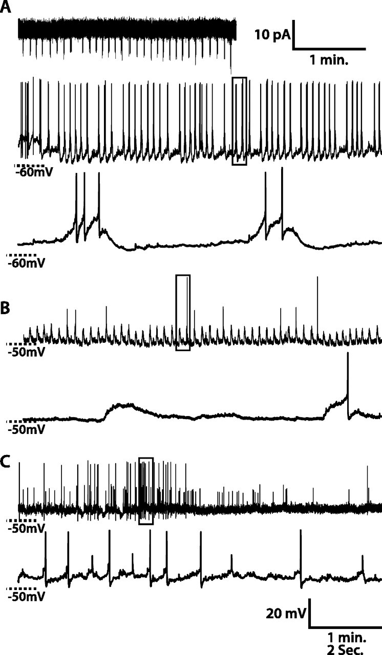
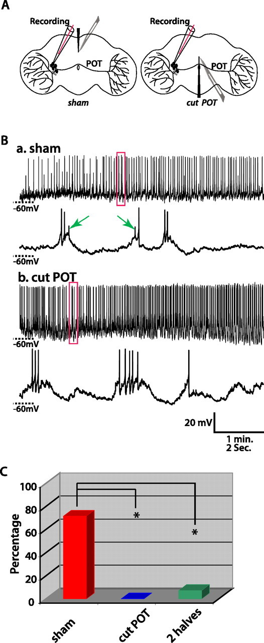
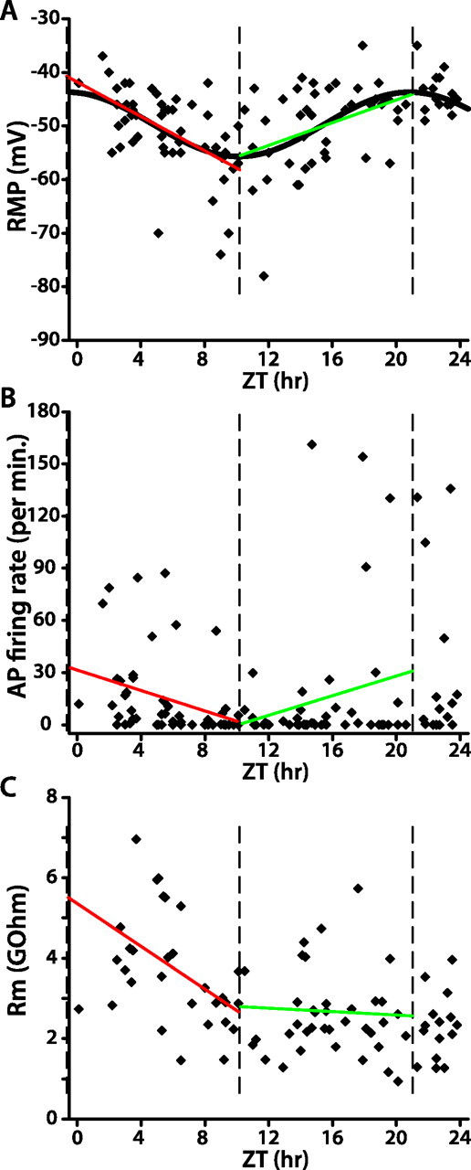
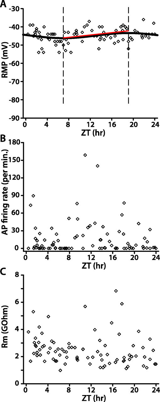
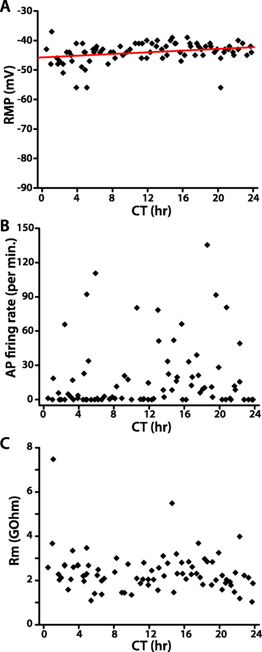
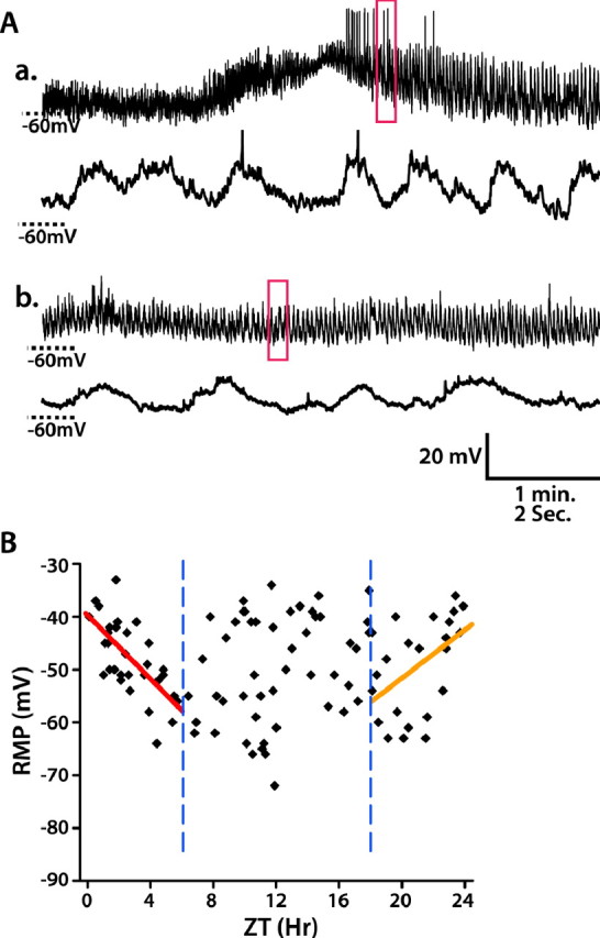
Drosophila circadian rhythms are controlled by a neural circuit containing approximately 150 clock neurons. Although much is known about mechanisms of autonomous cellular oscillation, the connection between cellular oscillation and functional outputs that control physiological and behavioral rhythms is poorly understood. To address this issue, we performed whole-cell patch-clamp recordings on lateral ventral clock neurons (LN(v)s), including large (lLN(v)s) and small LN(v)s (sLN(v)s), in situ in adult fly whole-brain explants. We found two distinct sizes of action potentials (APs) in >50% of lLN(v)s that fire APs spontaneously, and determined that large APs originate in the ipsilateral optic lobe and small APs in the contralateral. lLN(v) resting membrane potential (RMP), spontaneous AP firing rate, and membrane resistance are cyclically regulated as a function of time of day in 12 h light/dark conditions (LD). lLN(v) RMP becomes more hyperpolarized as time progresses from dawn to dusk with a concomitant decrease in spontaneous AP firing rate and membrane resistance. From dusk to dawn, lLN(v) RMP becomes more depolarized, with spontaneous AP firing rate and membrane resistance remaining stable. In contrast, circadian defective per(0) null mutant lLN(v) membrane excitability is nearly constant in LD. Over 24 h in constant darkness (DD), wild-type lLN(v) membrane excitability is not cyclically regulated, although RMP gradually becomes slightly more depolarized. sLN(v) RMP is most depolarized around lights-on, with substantial variability centered around lights-off in LD. Our results indicate that LN(v) membrane excitability encodes time of day via a circadian clock-dependent mechanism, and likely plays a critical role in regulating Drosophila circadian behavior.


Figures

Figure 1.
Figure 1.
Spontaneous activity of WT lLNv clock neurons. A–C, Three examples of whole-cell recordings from lLNvs. A, Top, A 3 min recording in cell-attached configuration. The middle panel in A and top panels in B and C are 5 min recordings in whole-cell current-clamp configuration. The bottom panels depict magnified views of the regions of the respective 5 min recordings outlined in the frame. For the lLNv in A, spontaneous AP firing rates and patterns are similar in cell-attached and in whole-cell configurations. lLNv RMP oscillates, and bursts of spontaneous APs occur on the depolarized phase of the RMP oscillations. For the lLNv in B, RMP oscillates, and spontaneous APs occur on the depolarized phase of some RMP oscillations but not the majority of them. For the lLNv in C, RMP does not oscillate and two different sizes of APs occur.
Figure 2.
Figure 2.
Gap junction inhibitors do not affect the amplitude of small APs. One example showing the effect of gap junction inhibitor, halothane (0.1%), on APs in lLNvs is shown. The lLNv is incubated in external solution containing 0.1% halothane for 20 min before washout with normal external solution. Several 1 min traces before and after the application of halothane and after washout are shown here. The frequency but not amplitude of small APs decreases after 10 min treatment of halothane.
Figure 3.
Figure 3.
Small APs of lLNvs originate in the contralateral optic lobe. A, Schematic diagrams of experimental design to address the origin of small APs. The left diagram shows the sham operation, in which a cut is made at the dorsal aspect of the brain without damaging the POT. The right diagram shows the experimental operation, in which the POT is cut. B, Representative traces recorded in the control and experimental group. In the control (a), the sham operation does not eliminate small APs (indicated by arrows). In the experimental group (b), only large APs occur. C, Histogram summarizing the percentage of lLNvs that fire both small and large APs after sham operation (red), cutting the POT (blue), and completely separating the brain into two halves (green). One silent cell that did not fire any APs was excluded from this analysis. The sham operation group was significantly different from both experimental groups, cutting the POT, or completely separating two hemispheres (χ2 test, both *p < 0.005).
Figure 4.
Figure 4.
WT lLNv membrane excitability in LD is strongly rhythmic. A–C, Each data point in the scatter plots represents the RMP (A), spontaneous AP firing rate (B), or membrane resistance (C) of one lLNv at the indicated time of recording. In A, the sinusoidal curve represents the best fit of RMP as a function of time of day in LD (ZT, nonlinear regression; p < 0.0001). The three vertical dashed lines indicate the peak and trough of the fitted sinusoid of RMP versus ZT. Red and green lines depict the linear regression analysis of data within those two intervals (A, both red and green linear fits, p < 0.01; B, C, red linear fit, p < 0.05; green linear fit, p > 0.1).
Figure 5.
Figure 5.
per0 lLNv membrane excitability in LD is only very weakly rhythmic. A–C, Scatter plots of per0 lLNv RMP (A), spontaneous AP firing rate (B), and membrane resistance (C). In A, the sinusoid represents the best fit of RMP versus time of day (nonlinear regression analysis, p < 0.0001), two vertical dashed lines depict the trough and peak of the sinusoidal curve, and the red line is the linear regression analysis of the data between the trough and peak (p > 0.1).
Figure 6.
Figure 6.
WT lLNv membrane excitability in DD is not rhythmic. A–C, Scatter plots of WT lLNv RMP (A), spontaneous AP firing rate (B), and membrane resistance (C). In A, the red line depicts the linear regression analysis of the relationship between RMP and time of day (p < 0.05).
Figure 7.
Figure 7.
WT sLNv RMP is most depolarized near lights-on. A, Examples of whole-cell recordings on sLNvs. a, b, Top panels are 5 min recordings, and bottom panels are the enlargement of the 10 s traces indicated in the red box of 5 min traces. a, An example of an sLNv that exhibits spontaneous APs. b, An example of an sLNv that exhibits no APs. B, Scatter plot of WT sLNv RMP versus time of day in LD. The relationship between RMP and time of day is analyzed using linear regression analysis. From ZT0 to ZT6, RMP becomes hyperpolarized (red line, p < 0.0001), and from ZT18 to ZT24 it becomes depolarized (orange line, p < 0.05), thus indicating a peak depolarization around lights-on. Two blue vertical dashed lines mark ZT6 and ZT18, respectively.

References

    1. Colwell CS. Rhythmic coupling among cells in the suprachiasmatic nucleus. J Neurobiol. 2000;43:379–388. - PMC - PubMed
    1. de Jeu M, Hermes M, Pennartz C. Circadian modulation of membrane properties in slices of rat suprachiasmatic nucleus. NeuroReport. 1998;9:3725–3729. - PubMed
    1. Green DJ, Gillette R. Circadian rhythm of firing rate recorded from single cells in the rat suprachiasmatic brain slice. Brain Res. 1982;245:198–200. - PubMed
    1. Grima B, Chelot E, Xia R, Rouyer F. Morning and evening peaks of activity rely on different clock neurons of the Drosophila brain. Nature. 2004;431:869–873. - PubMed
    1. Groos G, Hendriks J. Circadian rhythms in electrical discharge of rat suprachiasmatic neurones recorded in vitro. Neurosci Lett. 1982;34:283–288. - PubMed

Publication types

MeSH terms

LinkOut - more resources