

Preservation of peritubular capillary endothelial integrity and increasing pericytes may be critical to recovery from postischemic acute kidney injury
- PMID: 18562634
- PMCID: PMC2519188
- DOI: 10.1152/ajprenal.90276.2008
Preservation of peritubular capillary endothelial integrity and increasing pericytes may be critical to recovery from postischemic acute kidney injury
Abstract
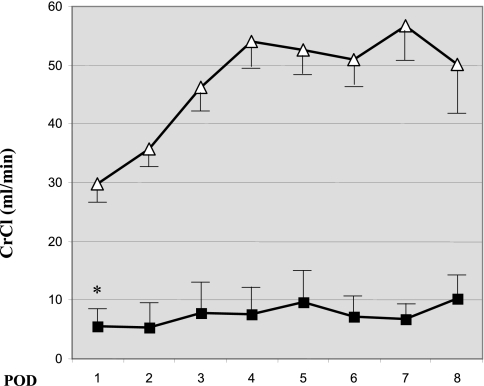
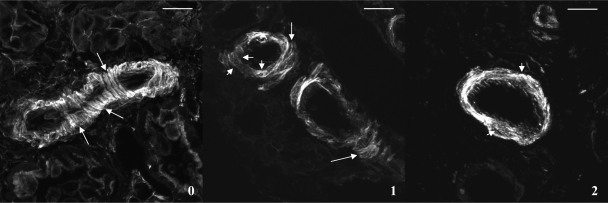
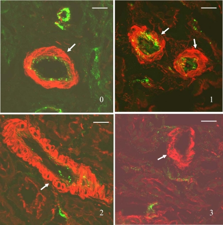
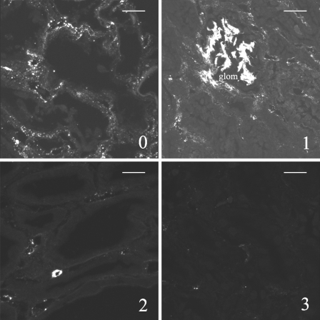
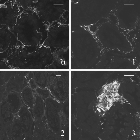
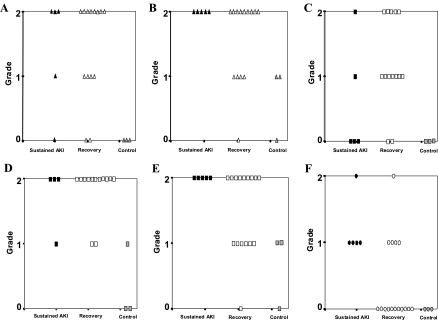
Decreased renal blood flow following an ischemic insult contributes to a reduction in glomerular filtration. However, little is known about the underlying cellular or subcellular mechanisms mediating reduced renal blood flow in human ischemic acute kidney injury (AKI) or acute renal failure (ARF). To examine renal vascular injury following ischemia, intraoperative graft biopsies were performed after reperfusion in 21 cadaveric renal allografts. Confocal fluorescence microscopy was utilized to examine vascular smooth muscle and endothelial cell integrity as well as peritubular interstitial pericytes in the biopsies. The reperfused, transplanted kidneys exhibited postischemic injury to the renal vasculature, as demonstrated by disorganization/disarray of the actin cytoskeleton in vascular smooth muscle cells and disappearance of von Willebrand factor from vascular endothelial cells. Damage to peritubular capillary endothelial cells was more severe in subjects destined to have sustained ARF than in those with rapid recovery of their graft function. In addition, peritubular pericytes/myofibroblasts were more pronounced in recipients destined to recover than those with sustained ARF. Taken together, these data suggest damage to the renal vasculature occurs after ischemia-reperfusion in human kidneys. Preservation of peritubular capillary endothelial integrity and increasing pericytes may be critical to recovery from postischemic AKI.
Figures
Similar articles
-
Ischemia induces alterations in actin filaments in renal vascular smooth muscle cells.Am J Physiol Renal Physiol. 2002 Jun;282(6):F1012-9. doi: 10.1152/ajprenal.00294.2001. Am J Physiol Renal Physiol. 2002. PMID: 11997317
-
Diminished NO generation by injured endothelium and loss of macula densa nNOS may contribute to sustained acute kidney injury after ischemia-reperfusion.Am J Physiol Renal Physiol. 2009 Jan;296(1):F25-33. doi: 10.1152/ajprenal.90531.2008. Epub 2008 Oct 29. Am J Physiol Renal Physiol. 2009. PMID: 18971208
-
Bone marrow-derived endothelial progenitor cells and endothelial cells may contribute to endothelial repair in the kidney immediately after ischemia-reperfusion.J Histochem Cytochem. 2010 Aug;58(8):687-94. doi: 10.1369/jhc.2010.956011. Epub 2010 Mar 30. J Histochem Cytochem. 2010. PMID: 20354148 Free PMC article.
-
The endothelial cell in ischemic acute kidney injury: implications for acute and chronic function.Kidney Int. 2007 Jul;72(2):151-6. doi: 10.1038/sj.ki.5002312. Epub 2007 May 2. Kidney Int. 2007. PMID: 17495858 Review.
-
Molecular mechanisms of acute renal failure following ischemia/reperfusion.Int J Artif Organs. 2004 Dec;27(12):1019-29. doi: 10.1177/039139880402701203. Int J Artif Organs. 2004. PMID: 15645611 Review.
Cited by
-
Recent advances in molecular mechanisms of acute kidney injury in patients with diabetes mellitus.Front Endocrinol (Lausanne). 2023 Jan 5;13:903970. doi: 10.3389/fendo.2022.903970. eCollection 2022. Front Endocrinol (Lausanne). 2023. PMID: 36686462 Free PMC article. Review.
-
Tipping the balance from angiogenesis to fibrosis in CKD.Kidney Int Suppl (2011). 2014 Nov;4(1):45-52. doi: 10.1038/kisup.2014.9. Kidney Int Suppl (2011). 2014. PMID: 26312149 Free PMC article. Review.
-
The Fibrin Cleavage Product Bβ15-42 Channels Endothelial and Tubular Regeneration in the Post-acute Course During Murine Renal Ischemia Reperfusion Injury.Front Pharmacol. 2018 Apr 27;9:369. doi: 10.3389/fphar.2018.00369. eCollection 2018. Front Pharmacol. 2018. PMID: 29755348 Free PMC article.
-
Ischemia and Reperfusion Injury in Kidney Transplantation: Relevant Mechanisms in Injury and Repair.J Clin Med. 2020 Jan 17;9(1):253. doi: 10.3390/jcm9010253. J Clin Med. 2020. PMID: 31963521 Free PMC article. Review.
-
Renal macro- and microcirculation autoregulatory capacity during early sepsis and norepinephrine infusion in rats.Crit Care. 2013 Jul 12;17(4):R139. doi: 10.1186/cc12818. Crit Care. 2013. PMID: 23849307 Free PMC article.
References
-
- Ali T, Khan I, Simpson W, Prescott G, Townend J, Smith W, MacLeod A. Incidence and outcomes in acute kidney injury: a comprehensive population-based study. J Am Soc Nephrol 18: 1292–1298, 2007. - PubMed
-
- Arendhorst WJ, Finn WF, Gottschalk CW. Micropuncture study of acute renal failure following temporary renal ischemia in the rat. Kidney Int 10, Suppl 6: S100–S105, 1976. - PubMed
-
- Basile DP, Donohoe DL, Roethe K, Mattson DL. Chronic renal hypoxia after acute ischemic injury: effects of l-arginine on hypoxia and secondary damage. Am J Physiol Renal Physiol 284: F338–F348, 2003. - PubMed
-
- Borawski J, Naumnik B, Pawlak K, Mysliwiec M. Endothelial dysfunction marker von Willebrand factor antigen in hemodialysis patients: association with pre-dialysis blood pressure and the acute phase response. Nephrol Dial Transplant 16: 1442–1447, 2001. - PubMed
Publication types
MeSH terms
Substances
Grants and funding
LinkOut - more resources
Full Text Sources
Other Literature Sources
Medical