. 2008 Jul;179(3):1221-35.
doi: 10.1534/genetics.107.083493. Epub 2008 Jun 18.

Meiotic recombination at the ends of chromosomes in Saccharomyces cerevisiae

Affiliations

Meiotic recombination at the ends of chromosomes in Saccharomyces cerevisiae

Arnold B Barton et al. Genetics. 2008 Jul.

Abstract

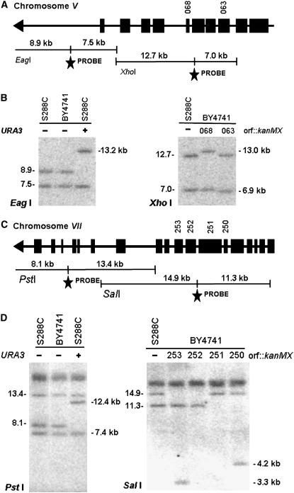
Meiotic reciprocal recombination (crossing over) was examined in the outermost 60-80 kb of almost all Saccharomyces cerevisiae chromosomes. These sequences included both repetitive gene-poor subtelomeric heterochromatin-like regions and their adjacent unique gene-rich euchromatin-like regions. Subtelomeric sequences underwent very little crossing over, exhibiting approximately two- to threefold fewer crossovers per kilobase of DNA than the genomic average. Surprisingly, the adjacent euchromatic regions underwent crossing over at twice the average genomic rate and contained at least nine new recombination "hot spots." These results prompted an analysis of existing genetic mapping data, which showed that meiotic reciprocal recombination rates were on average greater near chromosome ends exclusive of the subtelomeres. Thus, the distribution of crossovers in S. cerevisiae appears to resemble that found in several higher eukaryotes where the outermost chromosomal regions show increased crossing over.


Figures

F<sc>igure</sc> 1.—
Figure 1.—
Genetic mapping near chromosome ends. The amount of recombination (in centimorgans) between the telomeric URA3 and each kanMX insert in a nonessential gene was determined by tetrad analysis. The amount of recombination between the two kanMX markers (distance C) was estimated by subtracting distance A from distance B. ORFs are represented by solid boxes and telomeres by arrowheads. The structure of the ∼5-kb plasmid pEL61 used to mark all telomeres that was integrated into the C(1–3)A telomeric repeats (Louis and Borts 1995) is shown. Vector sequences are denoted by the hatched lines and telomeric repeat sequences by the arrowheads.
F<sc>igure</sc> 2.—
Figure 2.—
Recombination within the endmost regions of each chromosome. Recombination rates (centimorgans/kilobases) for 31 of 32 chromosome ends were determined as described in materials and methods and Figure 1. The locations of open reading frames are indicated below each panel. Subtelomeric regions are identified as either telomere-linked repetitive sequences or regions with a low density of ORFs. Repetitive sequences were defined as those showing >50% nucleic acid sequence identity to at least one other region of the genome (http//www.nottingham.ac.uk/biology/contact/academics/louis/ClustersSmall.html; SGD; Cherry et al. 1997). Low ORF density was arbitrarily defined as sequences that contained <60% “non-dubious” ORFs (as listed in SGD) using a 20-kb sliding window. ND, not determined. For chromosome IL, all displayed data but the leftmost interval are from prior studies that included the intervals iHIS3-iTRP1-23, iTRP1-23-pURA3, and pURA3-pyk1 (Su et al. 2000).
F<sc>igure</sc> 3.—
Figure 3.—
Effects of hemizygous insertions on reciprocal recombination. Physical maps showing the location of genes and ORFs replaced by kanMX or NAT1. The amounts of recombination (in centimorgans ± SE) between indicated markers are shown. Tetrad data not listed in Table 1 are shown in parentheses (PD:NPD:T). (A) Recombination on chromosome VIIL. (B) Recombination on chromosome VIR. (C) Recombination between ADE1(YAR015) and the telomeric pho11∷LEU2 on chromosome IR.
F<sc>igure</sc> 4.—
Figure 4.—
Increased meiotic reciprocal recombination near chromosome ends. Recombination rates (centimorgan/kilobase) of small- to medium-size physical intervals (2–100 kb ORF center to ORF center distance) were determined from existing SGD data and plotted as a function of (A) physical distance (kilobases) or (B) relative distance (fraction of total chromosome length) of the end of the interval to the telomere. Solid diamonds and solid linear regression trend lines represent SGD data. Open squares show all nonsubtelomeric data from this study. Regions were excluded as subtelomeric if they were both repetitive and had a low density of genes as defined in Figure 2. Dashed linear regression trend lines represent SGD data combined with data from this study. (C) Recombination rates decrease with increasing distance from the end of the chromosome. The slopes ±SE of the linear regressions measuring recombination rates vs. physical distance from the telomere were determined at the noted included distances from the telomere. All data are from supplemental Tables S1 and S2 and Table 1.
F<sc>igure</sc> 5.—
Figure 5.—
Gene conversion of kanMX as a function of chromosome position. Percentage of asci showing 3:1 and 1:3 segregation for each kanMX insert vs. its distance from the telomere (kilobases) are shown (see also Table 1). Solid diamonds denote kanMX markers that are adjacent to new reciprocal recombination hot spots.
F<sc>igure</sc> 6.—
Figure 6.—
Physical localization of URA3 and kanMX markers. (A) Physical map of the left end of chromosome V showing fragment sizes generated by noted endonucleases and locations of PCR-generated probes (stars). Arrowheads indicate telomeres and solid boxes indicate ORFs. Numbers above solid boxes indicate the location of corresponding ORF∷ kanMX inserts. The length of the inserted URA3 telomere marker is 4.3 kb (Louis and Borts 1995). (B) Blot hybridization showing noted endonuclease-digested genomic DNAs from parent haploid strains S288C and BY4741 containing specific markers on chromosome V. (+) and (−) indicate presence or absence of specific telomeric URA3 inserts and numbers correspond to the kanMX marked ORF. (C) Physical map of the left end of chromosome VII showing fragment sizes generated by noted endonucleases and locations of PCR-generated probes. Notations are as in A. (D) Blot hybridization showing noted endonuclease-digested genomic DNAs from chromosome VII marked strains. Notations are as in B. The kanMX insert introduces a SalI site resulting in shorter SalI fragments. The smallest fragments were run off the gel to allow resolution of the large bands. Additional bands that show no change are due to repetitive sequences located elsewhere in the genome.

Similar articles

Cited by

References

    1. Barlow, A. L., and M. A. Hulten, 1998. Crossing over analysis at pachytene in man. Eur. J. Hum. Genet. 6 350–358. - PubMed
    1. Barton, A. B., Y. Su, J. Lamb, D. Barber and D. B. Kaback, 2003. A function for subtelomeric DNA in Saccharomyces cerevisiae. Genetics 165 929–934. - PMC - PubMed
    1. Baudat, F., and A. Nicolas, 1997. Clustering of meiotic double-stranded breaks on yeast chromosome III. Proc. Natl. Acad. Sci. USA 94 5213–5218. - PMC - PubMed
    1. Blitzblau, H. G., G. W. Bell, J. Rodriquez, S. P. Bell and A. Hochwagen, 2007. Mapping of meiotic single-stranded DNA reveals double-strand break hotspots near telomeres and centromeres. Curr. Biol. 17 2003–2012. - PubMed
    1. Buhler, C., V. Borde and M. Lichten, 2007. Mapping meiotic single-strand DNA reveals a new landscape of DNA double-strand breaks in Saccharomyces cerevisiae. PLoS Biol. 5 2797–2808. - PMC - PubMed

Publication types

Substances