

Long noncoding RNAs in mouse embryonic stem cell pluripotency and differentiation
- PMID: 18562676
- PMCID: PMC2527704
- DOI: 10.1101/gr.078378.108
Long noncoding RNAs in mouse embryonic stem cell pluripotency and differentiation
Abstract
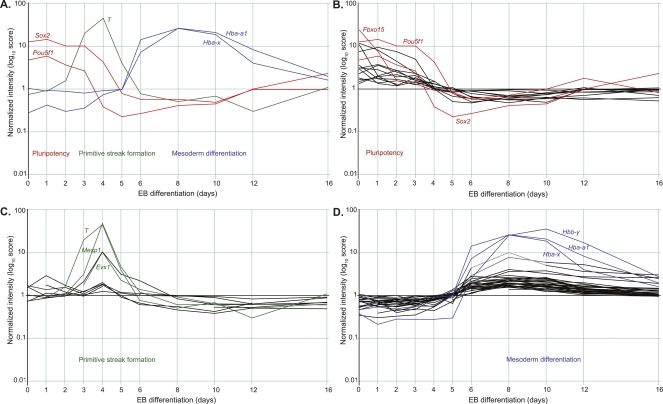
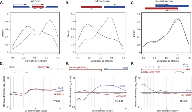
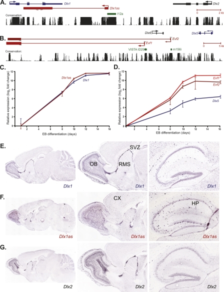
The transcriptional networks that regulate embryonic stem (ES) cell pluripotency and lineage specification are the subject of considerable attention. To date such studies have focused almost exclusively on protein-coding transcripts. However, recent transcriptome analyses show that the mammalian genome contains thousands of long noncoding RNAs (ncRNAs), many of which appear to be expressed in a developmentally regulated manner. The functions of these remain untested. To identify ncRNAs involved in ES cell biology, we used a custom-designed microarray to examine the expression profiles of mouse ES cells differentiating as embryoid bodies (EBs) over a 16-d time course. We identified 945 ncRNAs expressed during EB differentiation, of which 174 were differentially expressed, many correlating with pluripotency or specific differentiation events. Candidate ncRNAs were identified for further characterization by an integrated examination of expression profiles, genomic context, chromatin state, and promoter analysis. Many ncRNAs showed coordinated expression with genomically associated developmental genes, such as Dlx1, Dlx4, Gata6, and Ecsit. We examined two novel developmentally regulated ncRNAs, Evx1as and Hoxb5/6as, which are derived from homeotic loci and share similar expression patterns and localization in mouse embryos with their associated protein-coding genes. Using chromatin immunoprecipitation, we provide evidence that both ncRNAs are associated with trimethylated H3K4 histones and histone methyltransferase MLL1, suggesting a role in epigenetic regulation of homeotic loci during ES cell differentiation. Taken together, our data indicate that long ncRNAs are likely to be important in processes directing pluripotency and alternative differentiation programs, in some cases through engagement of the epigenetic machinery.
Figures

References
-
- Ball R.K., Friis R.R., Schoenenberger C.A., Doppler W., Groner B., Friis R.R., Schoenenberger C.A., Doppler W., Groner B., Schoenenberger C.A., Doppler W., Groner B., Doppler W., Groner B., Groner B. Prolactin regulation of beta-casein gene expression and of a cytosolic 120-kd protein in a cloned mouse mammary epithelial cell line. EMBO J. 1988;7:2089–2095. - PMC - PubMed
-
- Bejerano G., Pheasant M., Makunin I., Stephen S., Kent W.J., Mattick J.S., Haussler D., Pheasant M., Makunin I., Stephen S., Kent W.J., Mattick J.S., Haussler D., Makunin I., Stephen S., Kent W.J., Mattick J.S., Haussler D., Stephen S., Kent W.J., Mattick J.S., Haussler D., Kent W.J., Mattick J.S., Haussler D., Mattick J.S., Haussler D., Haussler D. Ultraconserved elements in the human genome. Science. 2004;304:1321–1325. - PubMed
-
- Bernstein E., Allis C.D., Allis C.D. RNA meets chromatin. Genes & Dev. 2005;19:1635–1655. - PubMed
-
- Bernstein B.E., Mikkelsen T.S., Xie X., Kamal M., Huebert D.J., Cuff J., Fry B., Meissner A., Wernig M., Plath K., Mikkelsen T.S., Xie X., Kamal M., Huebert D.J., Cuff J., Fry B., Meissner A., Wernig M., Plath K., Xie X., Kamal M., Huebert D.J., Cuff J., Fry B., Meissner A., Wernig M., Plath K., Kamal M., Huebert D.J., Cuff J., Fry B., Meissner A., Wernig M., Plath K., Huebert D.J., Cuff J., Fry B., Meissner A., Wernig M., Plath K., Cuff J., Fry B., Meissner A., Wernig M., Plath K., Fry B., Meissner A., Wernig M., Plath K., Meissner A., Wernig M., Plath K., Wernig M., Plath K., Plath K., et al. A bivalent chromatin structure marks key developmental genes in embryonic stem cells. Cell. 2006;125:315–326. - PubMed
-
- Boyer L.A., Lee T.I., Cole M.F., Johnstone S.E., Levine S.S., Zucker J.P., Guenther M.G., Kumar R.M., Murray H.L., Jenner R.G., Lee T.I., Cole M.F., Johnstone S.E., Levine S.S., Zucker J.P., Guenther M.G., Kumar R.M., Murray H.L., Jenner R.G., Cole M.F., Johnstone S.E., Levine S.S., Zucker J.P., Guenther M.G., Kumar R.M., Murray H.L., Jenner R.G., Johnstone S.E., Levine S.S., Zucker J.P., Guenther M.G., Kumar R.M., Murray H.L., Jenner R.G., Levine S.S., Zucker J.P., Guenther M.G., Kumar R.M., Murray H.L., Jenner R.G., Zucker J.P., Guenther M.G., Kumar R.M., Murray H.L., Jenner R.G., Guenther M.G., Kumar R.M., Murray H.L., Jenner R.G., Kumar R.M., Murray H.L., Jenner R.G., Murray H.L., Jenner R.G., Jenner R.G., et al. Core transcriptional regulatory circuitry in human embryonic stem cells. Cell. 2005;122:947–956. - PMC - PubMed
Publication types
MeSH terms
Substances
LinkOut - more resources
Full Text Sources
Other Literature Sources
Molecular Biology Databases