

Chronic kidney disease and bone fracture: a growing concern
- PMID: 18563052
- PMCID: PMC4139042
- DOI: 10.1038/ki.2008.264
Chronic kidney disease and bone fracture: a growing concern
Abstract
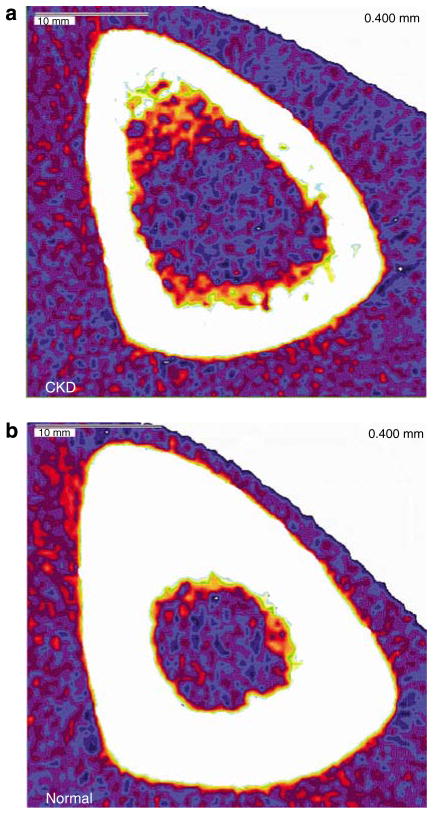
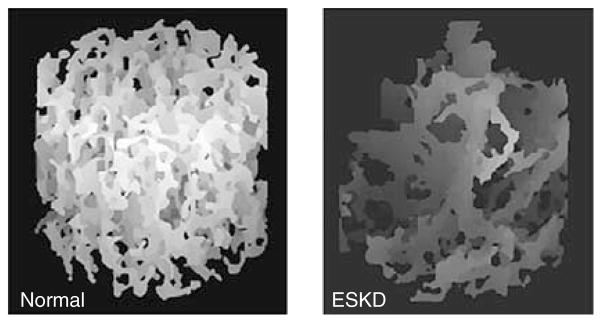
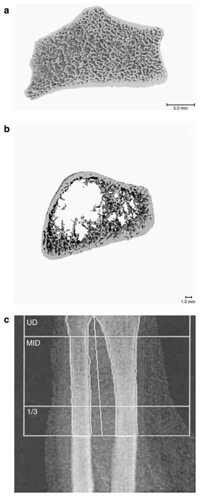
Susceptibility to fracture is increased across the spectrum of chronic kidney disease (CKD). Moreover, fracture in patients with end-stage kidney disease (ESKD) results in significant excess mortality. The incidence and prevalence of CKD and ESKD are predicted to increase markedly over the coming decades in conjunction with the aging of the population. Given the high prevalence of both osteoporosis and CKD in older adults, it is of the utmost public health relevance to be able to assess fracture risk in this population. Dual-energy X-ray absorptiometry (DXA), which provides an areal measurement of bone mineral density (aBMD), is the clinical standard to predict fracture in individuals with postmenopausal or age-related osteoporosis. Unfortunately, DXA does not discriminate fracture status in patients with ESKD. This may be, in part, because excess parathyroid hormone (PTH) secretion may accompany declining kidney function. Chronic exposure to high PTH levels preferentially causes cortical bone loss, which may be partially offset by periosteal expansion. DXA can neither reliably detect changes in bone volume nor distinguish between trabecular and cortical bone. In addition, DXA measurements may be low, normal, or high in each of the major forms of renal osteodystrophy (ROD). Moreover, postmenopausal or age-related osteoporosis may also affect patients with CKD and ESKD. Currently, transiliac crest bone biopsy is the gold standard to diagnose ROD and osteoporosis in patients with significant kidney dysfunction. However, bone biopsy is an invasive procedure that requires time-consuming analyses. Therefore, there is great interest in developing non-invasive high-resolution imaging techniques that can improve fracture risk prediction for patients with CKD. In this paper, we review studies of fracture risk in the setting of ESKD and CKD, the pathophysiology of increased fracture risk in patients with kidney dysfunction, the utility of various imaging modalities in predicting fracture across the spectrum of CKD, and studies evaluating the use of bisphosphonates in patients with CKD.
Figures
References
-
- Melton LJ., III Epidemiology worldwide. Endocrinol Metab Clin North Am. 2003;32:1–13. v. - PubMed
-
- Looker AC, Orwoll ES, Johnston CC, Jr, et al. Prevalence of low femoral bone density in older U.S. adults from III NHANES. J Bone Miner Res. 1997;12:1761–1768. - PubMed
-
- Cummings SR, Black DM, Nevitt MC, et al. Bone density at various sites for prediction of hip fractures. The Study of Osteoporotic Fractures Research Group. Lancet. 1993;341:72–75. - PubMed
-
- Cummings SR, Melton LJ. Epidemiology and outcomes of osteoporotic fractures. Lancet. 2002;359:1761–1767. - PubMed
-
- Burge R, Dawson-Hughes B, Solomon DH, et al. Incidence and economic burden of osteoporosis-related fractures in the United States, 2005–2025. J Bone Miner Res. 2007;22:465–475. - PubMed
Publication types
MeSH terms
Grants and funding
LinkOut - more resources
Full Text Sources
Other Literature Sources
Medical
