

Neuronal differentiation of synovial sarcoma and its therapeutic application
- PMID: 18563503
- PMCID: PMC2493002
- DOI: 10.1007/s11999-008-0343-z
Neuronal differentiation of synovial sarcoma and its therapeutic application
Abstract
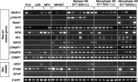
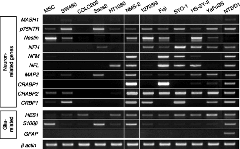
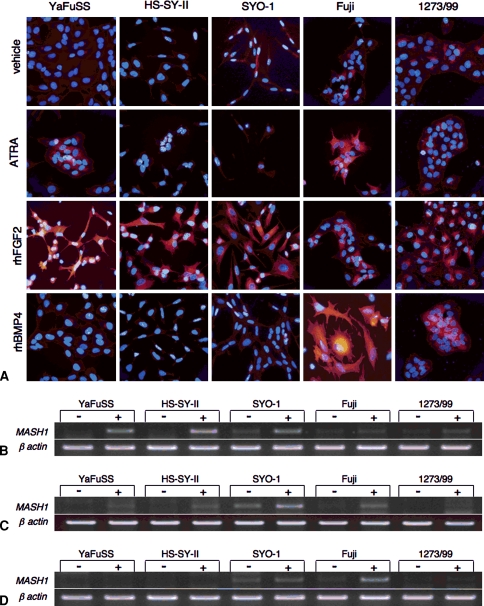
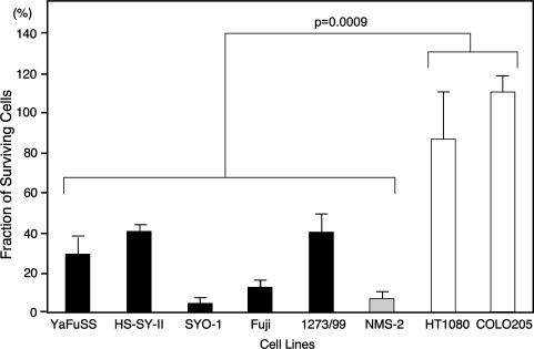
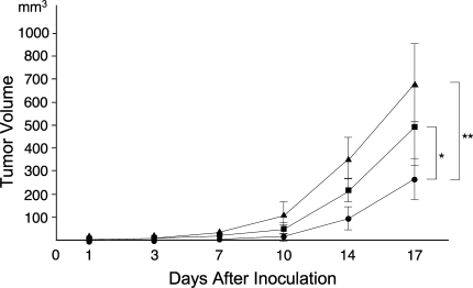
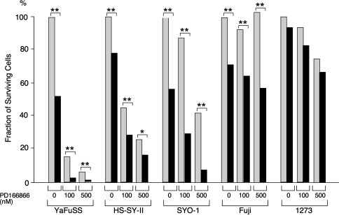
Synovial sarcoma is a rare sarcoma of unknown histologic origin. We previously reported the gene expression profile of synovial sarcoma was closely related to that of malignant peripheral nerve sheath tumors, and the fibroblast growth factor (FGF) signal was one of the main growth signals in synovial sarcoma. Here we further demonstrate the neural origin of synovial sarcoma using primary tumors and cell lines. The expression of neural tissue-related genes was confirmed in synovial sarcoma tumor tissues, but the expression of some genes was absent in synovial sarcoma cell lines. Treatment of synovial sarcoma cell lines with BMP4 or FGF2 enhanced or restored the expression of neural tissue-related genes and induced a neuron-like morphology with positive Tuj-1 expression. Treatment with all-trans-retinoic acid also induced the expression of neural tissue-related genes in association with growth inhibition, which was not observed in other cell lines except a malignant peripheral nerve sheath tumor cell line. A growth-inhibitory effect of all-trans-retinoic acid was also observed for xenografted tumors in athymic mice. The simultaneous treatment with FGF signal inhibitors enhanced the growth-inhibitory effect of all-trans-retinoic acid, suggesting the combination of growth signaling inhibition and differentiation induction could be a potential molecular target for treating synovial sarcoma.
Figures
Similar articles
-
Disruption of fibroblast growth factor signal pathway inhibits the growth of synovial sarcomas: potential application of signal inhibitors to molecular target therapy.Clin Cancer Res. 2005 Apr 1;11(7):2702-12. doi: 10.1158/1078-0432.CCR-04-2057. Clin Cancer Res. 2005. PMID: 15814652
-
Cluster analysis of immunohistochemical profiles in synovial sarcoma, malignant peripheral nerve sheath tumor, and Ewing sarcoma.Mod Pathol. 2006 May;19(5):659-68. doi: 10.1038/modpathol.3800569. Mod Pathol. 2006. PMID: 16528378
-
Neuronal differentiation of murine bone marrow Thy-1- and Sca-1-positive cells.J Hematother Stem Cell Res. 2003 Dec;12(6):727-34. doi: 10.1089/15258160360732740. J Hematother Stem Cell Res. 2003. PMID: 14977481
-
Synovial sarcoma of nerve.Hum Pathol. 2011 Apr;42(4):568-77. doi: 10.1016/j.humpath.2010.08.019. Epub 2011 Feb 4. Hum Pathol. 2011. PMID: 21295819 Review.
-
Of mice and men: opportunities to use genetically engineered mouse models of synovial sarcoma for preclinical cancer therapeutic evaluation.Cancer Control. 2011 Jul;18(3):196-203. doi: 10.1177/107327481101800307. Cancer Control. 2011. PMID: 21666582 Free PMC article. Review.
Cited by
-
Biphasic synovial sarcoma with a striking morphological divergence from the main mass to lymph node metastasis: A case report.Medicine (Baltimore). 2022 Jan 7;101(1):e28481. doi: 10.1097/MD.0000000000028481. Medicine (Baltimore). 2022. PMID: 35029897 Free PMC article.
-
Epigenetic Targets in Synovial Sarcoma: A Mini-Review.Front Oncol. 2019 Oct 18;9:1078. doi: 10.3389/fonc.2019.01078. eCollection 2019. Front Oncol. 2019. PMID: 31681608 Free PMC article. Review.
-
Synovial sarcoma: recent discoveries as a roadmap to new avenues for therapy.Cancer Discov. 2015 Feb;5(2):124-34. doi: 10.1158/2159-8290.CD-14-1246. Epub 2015 Jan 22. Cancer Discov. 2015. PMID: 25614489 Free PMC article. Review.
-
Functional Profiling of Soft Tissue Sarcoma Using Mechanistic Models.Int J Mol Sci. 2023 Sep 29;24(19):14732. doi: 10.3390/ijms241914732. Int J Mol Sci. 2023. PMID: 37834179 Free PMC article.
-
Reprogramming of mesenchymal stem cells by the synovial sarcoma-associated oncogene SYT-SSX2.Oncogene. 2012 May 3;31(18):2323-34. doi: 10.1038/onc.2011.418. Epub 2011 Sep 26. Oncogene. 2012. PMID: 21996728 Free PMC article.
References
-
- {'text': '', 'ref_index': 1, 'ids': [{'type': 'DOI', 'value': '10.1073/pnas.0530291100', 'is_inner': False, 'url': 'https://doi.org/10.1073/pnas.0530291100'}, {'type': 'PMC', 'value': 'PMC153034', 'is_inner': False, 'url': 'https://pmc.ncbi.nlm.nih.gov/articles/PMC153034/'}, {'type': 'PubMed', 'value': '12629218', 'is_inner': True, 'url': 'https://pubmed.ncbi.nlm.nih.gov/12629218/'}]}
- Al-Hajj M, Wicha MS, Benito-Hernandez A, Morrison SJ, Clarke MF. Prospective identification of tumorigenic breast cancer cells. Proc Natl Acad Sci USA. 2003;100:3983–3988. - PMC - PubMed
-
- {'text': '', 'ref_index': 1, 'ids': [{'type': 'PMC', 'value': 'PMC1850795', 'is_inner': False, 'url': 'https://pmc.ncbi.nlm.nih.gov/articles/PMC1850795/'}, {'type': 'PubMed', 'value': '12414507', 'is_inner': True, 'url': 'https://pubmed.ncbi.nlm.nih.gov/12414507/'}]}
- Allander SV, Illei PB, Chen Y, Antonescu CR, Bittner M, Ladanyi M, Meltzer PS. Expression profiling of synovial sarcoma by cDNA microarrays: association of ERBB2, IGFBP2, and ELF3 with epithelial differentiation. Am J Pathol. 2002;161:1587–1595. - PMC - PubMed
-
- {'text': '', 'ref_index': 1, 'ids': [{'type': 'PubMed', 'value': '6694356', 'is_inner': True, 'url': 'https://pubmed.ncbi.nlm.nih.gov/6694356/'}]}
- Andrews PW, Damjanov I, Simon D, Banting GS, Carlin C, Dracopoli NC, Fogh J. Pluripotent embryonal carcinoma clones derived from the human teratocarcinoma cell line Tera-2: differentiation in vivo and in vitro. Lab Invest. 1984;50:147–162. - PubMed
-
- {'text': '', 'ref_index': 1, 'ids': [{'type': 'DOI', 'value': '10.1097/00000478-200008000-00006', 'is_inner': False, 'url': 'https://doi.org/10.1097/00000478-200008000-00006'}, {'type': 'PubMed', 'value': '10935649', 'is_inner': True, 'url': 'https://pubmed.ncbi.nlm.nih.gov/10935649/'}]}
- Argani P, Faria PA, Epstein JI, Reuter VE, Perlman EJ, Beckwith JB, Ladanyi M. Primary renal synovial sarcoma: molecular and morphologic delineation of an entity previously included among embryonal sarcomas of the kidney. Am J Surg Pathol. 2000;24:1087–1096. - PubMed
-
- {'text': '', 'ref_index': 1, 'ids': [{'type': 'PubMed', 'value': '8314907', 'is_inner': True, 'url': 'https://pubmed.ncbi.nlm.nih.gov/8314907/'}]}
- Arnold HH, Braun T. The role of Myf-5 in somitogenesis and the development of skeletal muscles in vertebrates. J Cell Sci. 1993;104:957–960. - PubMed
Publication types
MeSH terms
Substances
LinkOut - more resources
Full Text Sources
