

Encapsulating a single G-quadruplex aptamer in a protein nanocavity
- PMID: 18563930
- PMCID: PMC2657931
- DOI: 10.1021/jp0775911
Encapsulating a single G-quadruplex aptamer in a protein nanocavity
Abstract
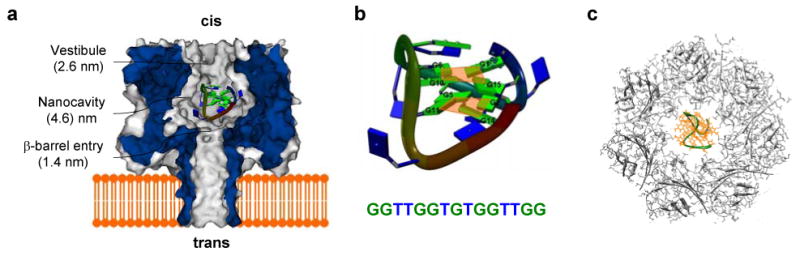
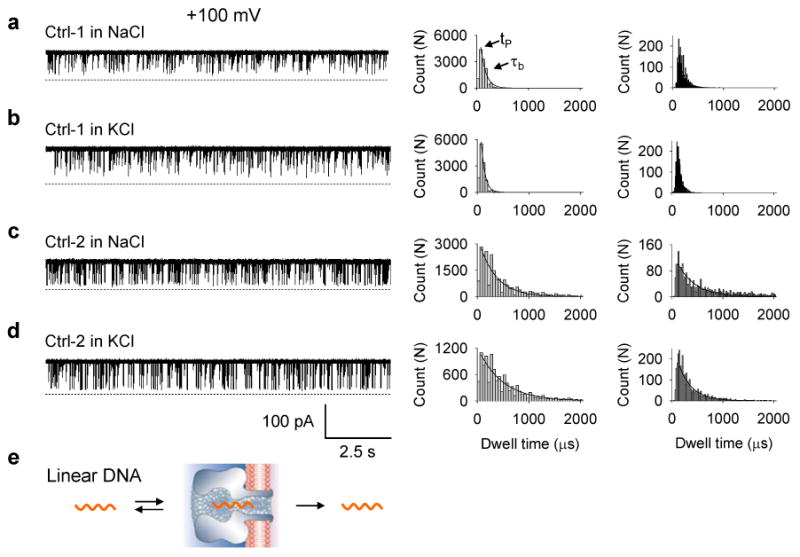
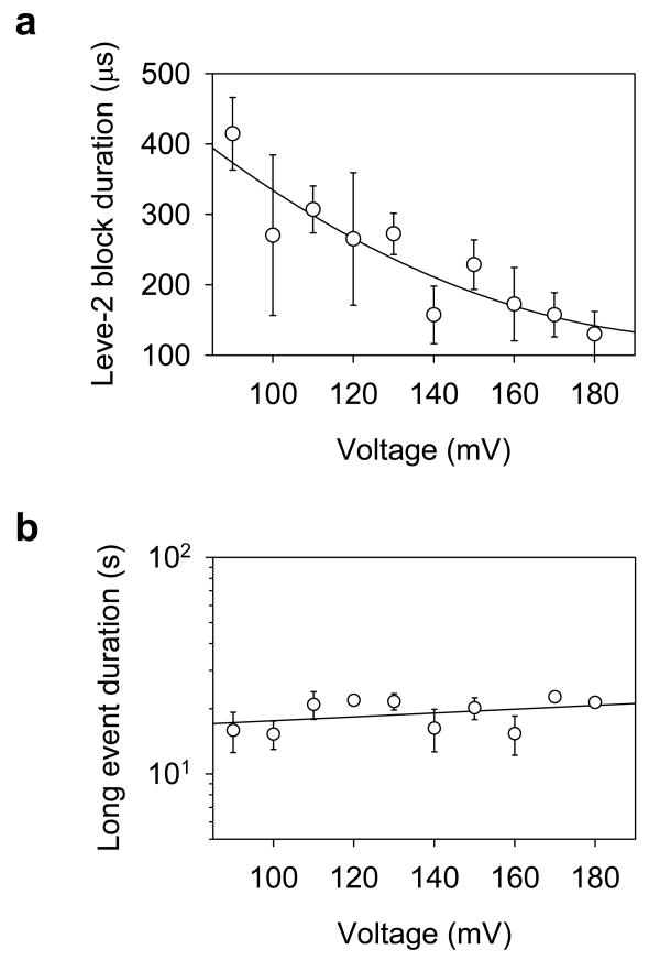
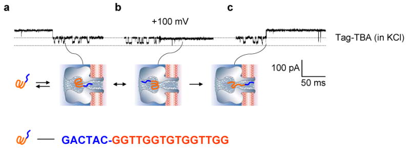
The alpha-hemolysin (alphaHL) protein pore has many applications in biotechnology. This article describes a single-molecule manipulation system that utilizes the nanocavity enclosed by this pore to noncovalently encapsulate a guest molecule. The guest is the thrombin-binding aptamer (TBA) that folds into the G-quadruplex in the presence of cations. Trapping the G-quadruplex in the nanocavity resulted in characteristic changes to the pore conductance that revealed important molecular processes, including spontaneous unfolding of the quartet structure and translocation of unfolded DNA in the pore. Through detection with Tag-TBA, we localized the G-quadruplex near the entry of the beta-barrel inside the nanocavity, where the molecule vibrates and rotates to different orientations. This guest-nanocavity supramolecular system has potential for helping to understand single-molecule folding and unfolding kinetics.
Figures

Similar articles
-
Single-molecule detection of folding and unfolding of the G-quadruplex aptamer in a nanopore nanocavity.Nucleic Acids Res. 2009 Feb;37(3):972-82. doi: 10.1093/nar/gkn968. Epub 2008 Dec 26. Nucleic Acids Res. 2009. PMID: 19112078 Free PMC article.
-
Single-molecule investigation of G-quadruplex folds of the human telomere sequence in a protein nanocavity.Proc Natl Acad Sci U S A. 2014 Oct 7;111(40):14325-31. doi: 10.1073/pnas.1415944111. Epub 2014 Sep 15. Proc Natl Acad Sci U S A. 2014. PMID: 25225404 Free PMC article.
-
Single-molecule investigation of G-quadruplex using a nanopore sensor.Methods. 2012 May;57(1):40-6. doi: 10.1016/j.ymeth.2012.03.026. Epub 2012 Apr 2. Methods. 2012. PMID: 22487183 Free PMC article. Review.
-
A DNA-Based Biosensor Assay for the Kinetic Characterization of Ion-Dependent Aptamer Folding and Protein Binding.Molecules. 2019 Aug 8;24(16):2877. doi: 10.3390/molecules24162877. Molecules. 2019. PMID: 31398834 Free PMC article.
-
Single molecule sensing by nanopores and nanopore devices.Analyst. 2010 Mar;135(3):441-51. doi: 10.1039/b907735a. Epub 2009 Dec 22. Analyst. 2010. PMID: 20174694 Free PMC article. Review.
Cited by
-
Beta-Barrel Nanopores as Diagnostic Sensors: An Engineering Perspective.Biosensors (Basel). 2024 Jul 16;14(7):345. doi: 10.3390/bios14070345. Biosensors (Basel). 2024. PMID: 39056622 Free PMC article. Review.
-
Quantitative analysis of the nanopore translocation dynamics of simple structured polynucleotides.Biophys J. 2012 Jan 4;102(1):85-95. doi: 10.1016/j.bpj.2011.11.4011. Epub 2012 Jan 3. Biophys J. 2012. PMID: 22225801 Free PMC article.
-
Biomedical diagnosis perspective of epigenetic detections using alpha-hemolysin nanopore.AIMS Mater Sci. 2015;2(4):448-472. doi: 10.3934/matersci.2015.4.448. Epub 2015 Nov 16. AIMS Mater Sci. 2015. PMID: 30931380 Free PMC article.
-
Probing molecular pathways for DNA orientational trapping, unzipping and translocation in nanopores by using a tunable overhang sensor.Nanoscale. 2014 Oct 7;6(19):11372-9. doi: 10.1039/c4nr03195d. Nanoscale. 2014. PMID: 25144935 Free PMC article.
-
Nanopore single-molecule dielectrophoretic detection of cancer-derived microRNA biomarkers.Annu Int Conf IEEE Eng Med Biol Soc. 2013;2013:6821-4. doi: 10.1109/EMBC.2013.6611124. Annu Int Conf IEEE Eng Med Biol Soc. 2013. PMID: 24111311 Free PMC article.
References
-
- Bayley H, Cremer PS. Nature. 2001;413:226–230. - PubMed
-
- Bayley H, Jayasinghe L. Molecular Membrane Biology. 2004;21:209–220. - PubMed
-
- Braha O, Gu LQ, Zhou L, Lu XF, Cheley S, Bayley H. Nature Biotechnology. 2000;18:1005–1007. - PubMed
-
- Gu LQ, Braha O, Conlan S, Cheley S, Bayley H. Nature. 1999;398:686–690. - PubMed
-
- Movileanu L, Howorka S, Braha O, Bayley H. Nature Biotechnology. 2000;18:1091–1095. - PubMed
Publication types
MeSH terms
Substances
Grants and funding
LinkOut - more resources
Full Text Sources
Other Literature Sources
