. 2008 Sep;9(9):1446-57.
doi: 10.1111/j.1600-0854.2008.00781.x. Epub 2008 Jun 28.

CD1a and MHC class I follow a similar endocytic recycling pathway

Affiliations

CD1a and MHC class I follow a similar endocytic recycling pathway

Duarte C Barral et al. Traffic. 2008 Sep.

Abstract

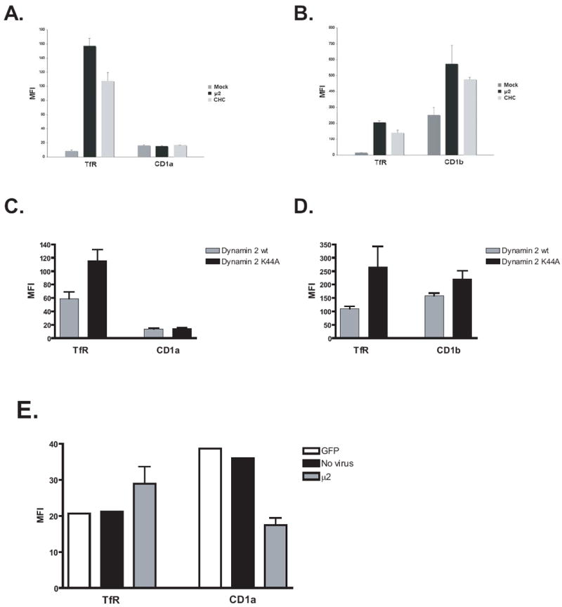
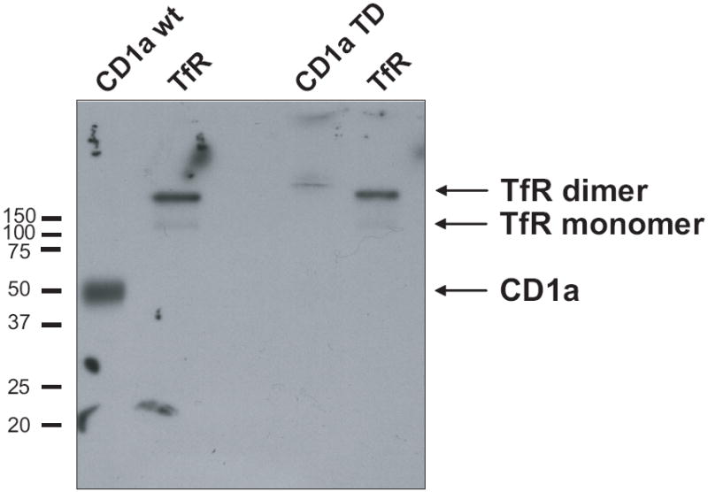
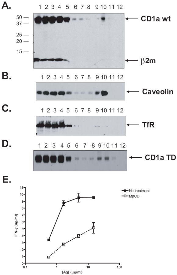
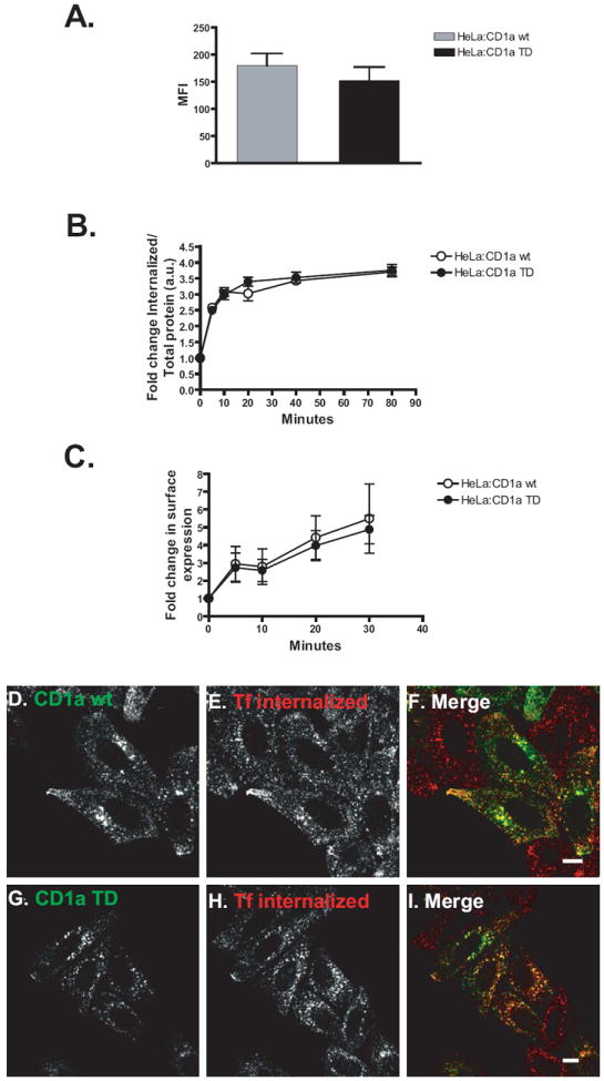
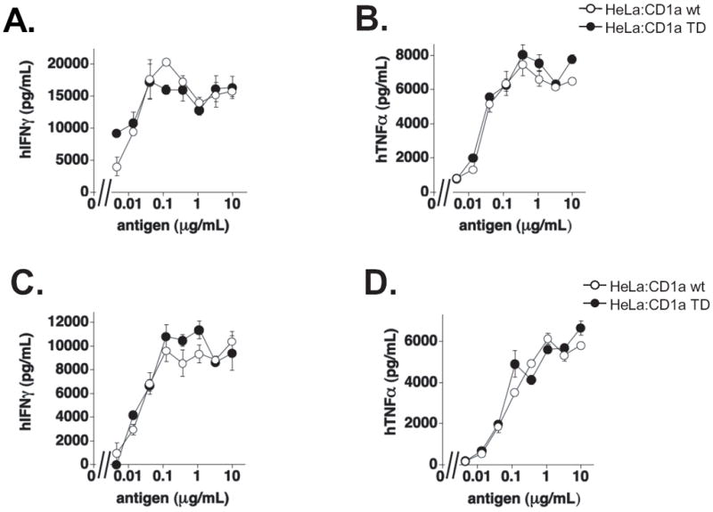
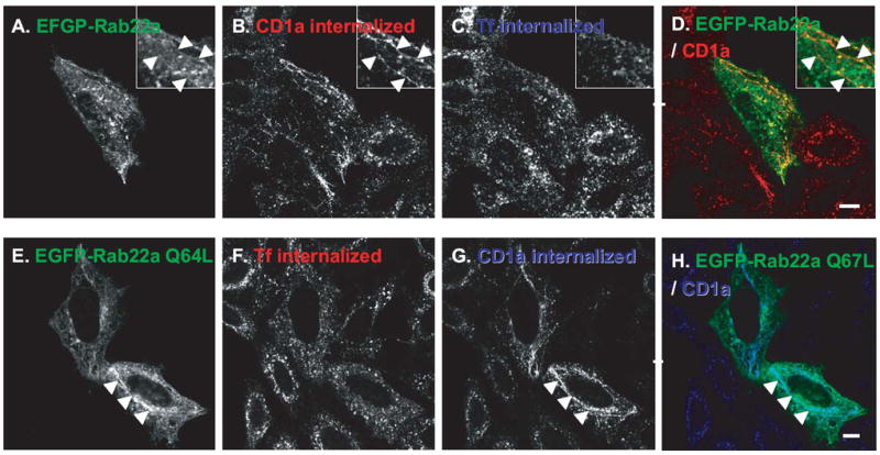
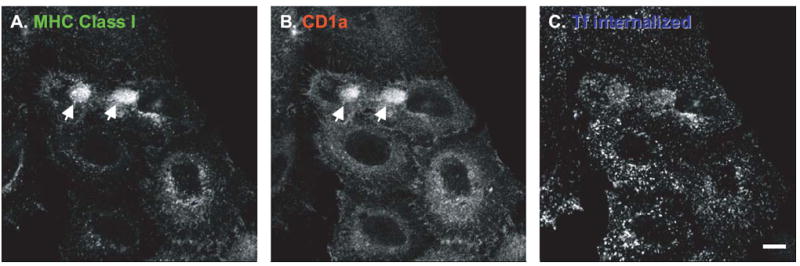
CD1 proteins are a family of major histocompatibility complex (MHC) class I-like antigen-presenting molecules that present lipids to T cells. The cytoplasmic tails (CTs) of all human CD1 isoforms, with the exception of CD1a, contain tyrosine-based sorting motifs, responsible for the internalization of proteins by the clathrin-mediated pathway. The role of the CD1a CT, which does not possess any sorting motifs, as well as its mode of internalization are not known. We investigated the internalization and recycling pathways followed by CD1a and the role of its CT. We found that CD1a can be internalized by a clathrin- and dynamin-independent pathway and that it follows a Rab22a- and ADP ribosylation factor (ARF)6-dependent recycling pathway, similar to other cargo internalized independent of clathrin. We also found that the CD1a CT is S-acylated. However, this posttranslational modification does not determine the rate of internalization or recycling of the protein or its localization to detergent-resistant membrane microdomains (DRMs) where we found CD1a to be enriched. We also show that plasma membrane DRMs are essential for efficient CD1a-mediated antigen presentation. These findings place CD1a closer to MHC class I in its trafficking and potential antigen-loading compartments among CD1 isoforms. Furthermore, we identify CD1a as a new marker for the clathrin- and dynamin-independent and DRM-dependent pathway of internalization as well as the Rab22a- and ARF6-dependent recycling pathway.


Figures

Figure 1
Figure 1. CD1a Surface Expression is Not Affected by Inhibition of the Clathrin-Mediated Pathway
HeLa:CD1a (Panels A and C) or HeLa:CD1b (Panels B and D) stable transfectants were transiently transfected with clathrin-heavy chain (CHC) or μ2-subunit of AP-2 (μ2) siRNA oligos, twice at 72 h intervals (Panels A and B), or with EGFP-Dynamin 2 wt or EGFP-Dynamin 2 K44A for 42 h (Panels C and D), then stained with anti-CD1a, anti-CD1b or anti-TfR antibodies and analyzed by flow cytometry. Error bars represent the standard deviation of three independent experiments. Panel E. CD14+ monocytes were differentiated into immature monocyte-derived dendritic cells and transduced (or not) with GFP or μ2 shRNA encoding lentivirus for 6 days and analyzed in the same way. The Mean Fluorescence Intensity (MFI) is represented in all cases and the error bars represent the standard deviation of four different shRNA for μ2.
Figure 2
Figure 2. CD1a wt, but Not CD1a TD, is Post-Translationally Modified by S-Acylation
HeLa:CD1a wt or Hela:CD1a TD stable transfectant cells were labeled overnight with 3H-palmitic acid and, after lysing the cells, the indicated proteins were immunoprecipitated with appropriate monoclonal antibodies. The immunoprecipitates were analyzed by 12.5% SDS-PAGE and the gels subjected to fluorography. The results are representative of two independent experiments.
Figure 3
Figure 3. CD1a Partitions to Detergent-Resistant Membrane Microdomains, which are Necessary for Efficient CD1a-Restricted Exogenous Antigen Presentation
HeLa:CD1a wt (Panel A) or HeLa:CD1a TD (Panel D) stable transfectant cells were surface biotinylated, lysed and fractionated in a sucrose step gradient. Fractions were collected from the bottom and CD1a immunoprecipitated (Panels A and D). Samples were resolved on 12.5% SDS-PAGE and immunoblotted with HRP-conjugated streptavidin (Panels A and D) or with rabbit polyclonal anti-caveolin (Panel B) or goat polyclonal anti-TfR (Panel C), followed by HRP-conjugated anti-rabbit or anti-goat antibodies, respectively. In Panel A, associated β2-microglobulin (β2m) is also shown. Results are representative of at least two independent experiments. Panel E. CD14+ monocytes from human donors were differentiated into immature monocyte-derived dendritic cells (DCs) and cholesterol-depleted by treatment with Methyl-β-cyclodextrin (MβCD). DCs were then fixed and incubated with indicated concentrations of sonicated antigen and CD8-2 T cells, which recognize dideoxymycobactin (DDM) presented by CD1a. After 21h, the release of IFN-γ was measured by ELISA. Error bars represent standard deviation of triplicate measurements and the results are representative of three independent experiments.
Figure 4
Figure 4. CD1a TD Shows Normal Surface Expression, Internalization and Recycling
Panel A. HeLa cells were transiently co-transfected with CD1a wt and EGFP or CD1a TD and EGFP, stained for CD1a and analyzed by flow cytometry, gating on EGFP+ cells. Panel B. HeLa:CD1a wt and HeLa:CD1a TD stable transfectants were surface biotinylated with a cleavable biotin at 4°C, shifted to 37°C for the indicated periods of time (to allow for internalization), after which cells were placed on ice to stop internalization. Surface biotin was cleaved with a reducing agent (on ice) and cells were then lysed and lysates probed for different proteins by ELISA with each condition performed in duplicate. The results (in arbitrary units) are normalized for the total CD1a protein present in each lysate and the internalization at time zero. Panel C. HeLa cells were transiently co-transfected with CD1a wt and EGFP or CD1a TD and EGFP, stained for CD1a and incubated at 37°C to allow for internalization. After stripping the surface-bound antibody with a brief acidic wash, cells were incubated at 37°C for different periods of time to allow for recycling. Cells were then stained with PE-labeled anti-mouse secondary antibody and analyzed by flow cytometry, gating on EGFP+ cells. The results (in arbitrary units) are normalized for the recycling at time zero. Error bars in panels A, B and C represent the standard deviation of three independent experiments. Panels D-I. HeLa cells were transiently transfected with CD1a wt (Panels D, E and F) or CD1a TD (Panels G, H and I), serum-starved for 30 minutes and incubated with anti-CD1a and Alexa 546-conjugated transferrin (Tf) on ice for 30 minutes. Cells were then washed and incubated with Alexa 546-conjugated Tf for 30 minutes at 37°C. After stripping the surface-bound antibody with a brief acidic wash, cells were fixed, permeabilized and labeled with Alexa 488-conjugated anti-mouse (Panels D and G) antibody. In the merge panels F and I, colocalization is indicated by the yellow color. Scale bars, 10 μm.
Figure 5
Figure 5. CD1a TD Efficiently Presents Sulfatide to T cells
Nervonoyl sulfatide, at the indicated concentrations, was incubated with HeLa:CD1a wt or HeLa:CD1a TD and the CD1a-restricted sulfatide-specific human T-cell clone K34 B9.1, during the whole assay (Panels A and B) or pulsed for 1 h at 37°C (Panels C and D). Supernatants were collected after 48 h and release of human IFN-γ (Panels A and C) and human TNF-α (Panels B and D) was measured by ELISA. Error bars represent the standard deviation of triplicate measurements.
Figure 6
Figure 6. CD1a Follows a Rab22a-Dependent Recycling Pathway
Hela:CD1a stable transfectants were transiently transfected with EGFP-Rab22a (wt or Q64L constitutively-active mutant). After 24 h, cells were serum-starved for 30 minutes and incubated with Alexa 647-conjugated transferrin (Tf, Panel C) or Alexa 546-conjugated Tf (Panel F) and anti-CD1a monoclonal antibody (Panels B and G), for 30 minutes on ice. Cells were then washed and incubated with Alexa 647-conjugated Tf for 45 minutes at 37°C (Upper panels) or switched only to 37°C for 30 minutes (Lower panels). After stripping the surface-bound antibody with a brief acidic wash, cells were fixed, permeabilized and labeled with Alexa 546-conjugated anti-mouse (Panel B) or Cy5-conjugated anti-mouse (Panel G) antibodies. Arrowheads depict tubular structures that contain both EGFP-Rab22a and CD1a. In the merge panel D, colocalization is indicated by the yellow color, while in panel H it is indicated by the color cyan. Scale bars, 10 μm.
Figure 7
Figure 7. CD1a Colocalizes with MHC Class I at Steady-State
HeLa:CD1a stable transfectant cells were fixed, permeabilized and stained with Alexa-488-conjugated anti-MHC class I (Panel A) and Alexa 546-conjugated anti-CD1a (Panel B) antibodies. In the merge panel, colocalization is indicated by the yellow color. Scale bars, 10 μm.
Figure 8
Figure 8. CD1a is Internalized and Recycled via an ARF6-Dependent Pathway
HeLa:CD1a stable transfectants were transiently transfected with ARF6-Q67L constitutively-active mutant. After 24 h, cells were serum starved for 30 minutes and incubated with Alexa 647-conjugated transferrin (Tf, Panel C) for 30 minutes on ice. Cells were washed, incubated with Alexa 647-conjugated Tf for 30 minutes at 37°C and then fixed, permeabilized and incubated with Alexa 488-conjugated anti-MHC class I (Panel A) and Alexa 546-conjugated anti-CD1a (Panel B) antibodies. Arrowheads depict typical enlarged vacuolar structures induced by ARF6-Q67L expression. Scale bar, 10 μm.

References

    1. Dascher CC. Evolutionary biology of CD1. Curr Top Microbiol Immunol. 2007;314:3–26. - PubMed
    1. Zajonc DM, Wilson IA. Architecture of CD1 proteins. Curr Top Microbiol Immunol. 2007;314:27–50. - PubMed
    1. Young DC, Moody DB. T-cell recognition of glycolipids presented by CD1 proteins. Glycobiology. 2006;16(7):103R–112R. - PubMed
    1. Mukherjee S, Soe TT, Maxfield FR. Endocytic sorting of lipid analogues differing solely in the chemistry of their hydrophobic tails. J Cell Biol. 1999;144(6):1271–1284. - PMC - PubMed
    1. Moody DB, Briken V, Cheng TY, Roura-Mir C, Guy MR, Geho DH, Tykocinski ML, Besra GS, Porcelli SA. Lipid length controls antigen entry into endosomal and nonendosomal pathways for CD1b presentation. Nat Immunol. 2002;3(5):435–442. - PubMed

Publication types

MeSH terms