. 2008 Jun 18:8:97.
doi: 10.1186/1471-2180-8-97.

Phagocytosis of Aspergillus fumigatus conidia by primary nasal epithelial cells in vitro

Affiliations

Phagocytosis of Aspergillus fumigatus conidia by primary nasal epithelial cells in vitro

Françoise Botterel et al. BMC Microbiol. .

Abstract

Background: Invasive aspergillosis, which is mainly caused by the fungus Aspergillus fumigatus, is an increasing problem in immunocompromised patients. Infection occurs by inhalation of airborne conidia, which are first encountered by airway epithelial cells. Internalization of these conidia into the epithelial cells could serve as a portal of entry for this pathogenic fungus.

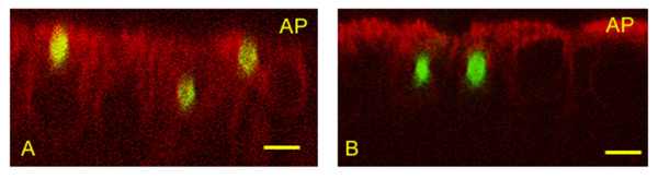
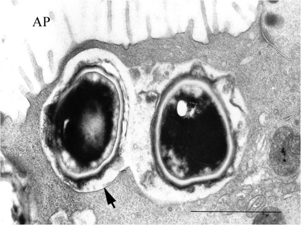
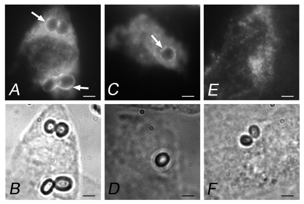
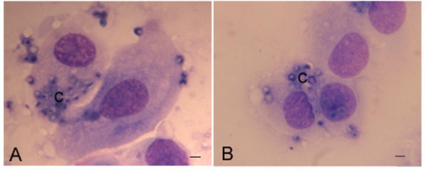
Results: We used an in vitro model of primary cultures of human nasal epithelial cells (HNEC) at an air-liquid interface. A. fumigatus conidia were compared to Penicillium chrysogenum conidia, a mould that is rarely responsible for invasive disease. Confocal microscopy, transmission electron microscopy, and anti-LAMP1 antibody labeling studies showed that conidia of both species were phagocytosed and trafficked into a late endosomal-lysosomal compartment as early as 4 h post-infection. In double immunolabeling experiments, the mean percentage of A. fumigatus conidia undergoing phagocytosis 4 h post-infection was 21.8 +/- 4.5%. Using combined staining with a fluorescence brightener and propidium iodide, the mean rate of phagocytosis was 18.7 +/- 9.3% and the killing rate 16.7 +/- 7.5% for A. fumigatus after 8 h. The phagocytosis rate did not differ between the two fungal species for a given primary culture. No germination of the conidia was observed until 20 h of observation.

Conclusion: HNEC can phagocytose fungal conidia but killing of phagocytosed conidia is low, although the spores do not germinate. This phagocytosis does not seem to be specific to A. fumigatus. Other immune cells or mechanisms are required to kill A. fumigatus conidia and to avoid further invasion.


Figures

Figure 1
Figure 1
Z view of HNEC showing internalization of A. fumigatus and P. chrysogenum conidia using confocal immunofluorescence microscopy. Cells were incubated with FITC-labeled conidia for 8 h. The cells were then labeled with specific human cytokeratin antibody and conjugated secondary antibody. (A) Three A. fumigatus conidia in HNEC. (B) Two P. chrysogenum conidia in HNEC. AP: apical pole. Bar = 5 μm.
Figure 2
Figure 2
Transmission electron microscopy of HNEC after 4 h incubation with A. fumigatus showing conidia internalized in a membrane-bound vacuole. Note the double-layered cell wall (arrow) and the electron-dense pigmented outer layer of the conidium. AP: apical pole. Bar = 1 μm.
Figure 3
Figure 3
Intracellular trafficking of A. fumigatus and P. chrysogenum conidia within HNEC phagosomes after 4 h incubation. After fixation and permeabilization, cells were labeled with specific mouse monoclonal anti-LAMP1 antibody (mAb) and secondary antibody conjugated to Texas-red. The lower figures are the corresponding phase contrast images. (A and B) A. fumigatus conidia; (C and D) P. chrysogenum conidia; (E and F) control HNEC labeled with mouse IgG1 mAb. Arrows indicate conidia stained positive for LAMP1. Bar = 2 μm.
Figure 4
Figure 4
HNEC stained with May-Grünwald Giemsa after 8 h incubation with A. fumigatus (A) and P. chrysogenum (B) conidia. Cells were released and smears prepared before staining. Quantification of HNEC and conidia was performed to determine the number of HNEC-associated conidia and the average number of conidia associated with HNEC. (c) = conidia. Bar = 2 μm.
Figure 5
Figure 5
Internalization of A. fumigatus conidia by HNEC. HNEC were incubated with FITC-labeled A. fumigatus conidia for 4 h, washed to remove unbound conidia, and fixed. HNEC were incubated with a rabbit anti-conidia antiserum and then with Texas-red-conjugated anti-rabbit antibody to label non-phagocytosed conidia. (A) Overlay of green and red channels; (B) red channel (non-phagocytosed conidia); (C) green channel (total conidia). Bar = 2 μm.

References

    1. Lin SJ, Schranz J, Teutsch SM. Aspergillosis case-fatality rate: systematic review of the literature. Clin Infect Dis. 2001;32:358–366. doi: 10.1086/318483. - DOI - PubMed
    1. Meersseman W, Lagrou K, Maertens J, Van Wijngaerden E. Invasive aspergillosis in the intensive care unit. Clin Infect Dis. 2007;45:205–216. doi: 10.1086/518852. - DOI - PubMed
    1. Singh N, Paterson DL. Aspergillus infections in transplant recipients. Clin Microbiol Rev. 2005;18:44–69. doi: 10.1128/CMR.18.1.44-69.2005. - DOI - PMC - PubMed
    1. Rhodes JC. Aspergillus fumigatus: growth and virulence. Med Mycol. 2006;44:S77–81. doi: 10.1080/13693780600779419. - DOI - PubMed
    1. Philippe B, Ibrahim-Granet O, Prevost MC, Gougerot-Pocidalo MA, Sanchez Perez M, Meeren A Van der, Latge JP. Killing of Aspergillus fumigatus by alveolar macrophages is mediated by reactive oxidant intermediates. Infect Immun. 2003;71:3034–3042. doi: 10.1128/IAI.71.6.3034-3042.2003. - DOI - PMC - PubMed

Publication types

LinkOut - more resources