Comparative Study
. 2008 Jun 18:9:293.
doi: 10.1186/1471-2164-9-293.

Genomic structure and expression of Jmjd6 and evolutionary analysis in the context of related JmjC domain containing proteins

Affiliations
Comparative Study

Genomic structure and expression of Jmjd6 and evolutionary analysis in the context of related JmjC domain containing proteins

Phillip Hahn et al. BMC Genomics. .

Abstract

Background: The jumonji C (JmjC) domain containing gene 6 (Jmjd6, previously known as phosphatidylserine receptor) has misleadingly been annotated to encode a transmembrane receptor for the engulfment of apoptotic cells. Given the importance of JmjC domain containing proteins in controlling a wide range of diverse biological functions, we undertook a comparative genomic analysis to gain further insights in Jmjd6 gene organisation, evolution, and protein function.

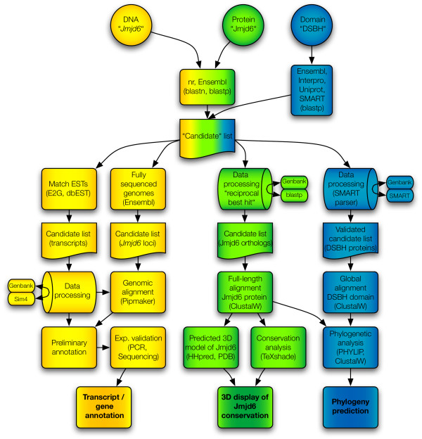
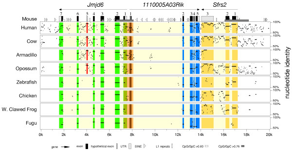
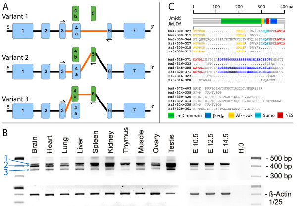
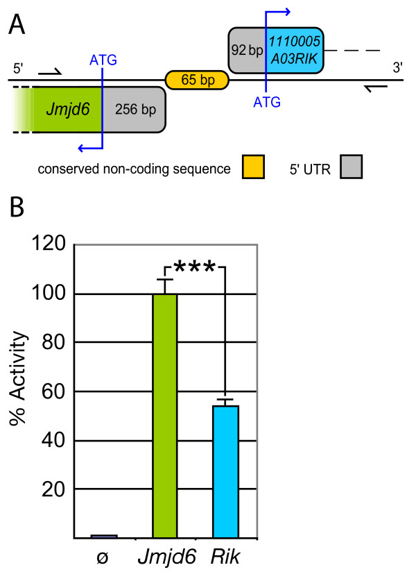
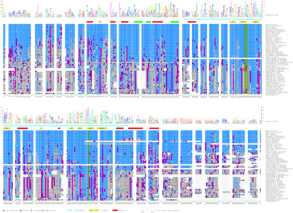
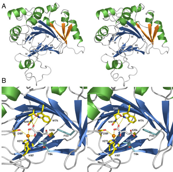
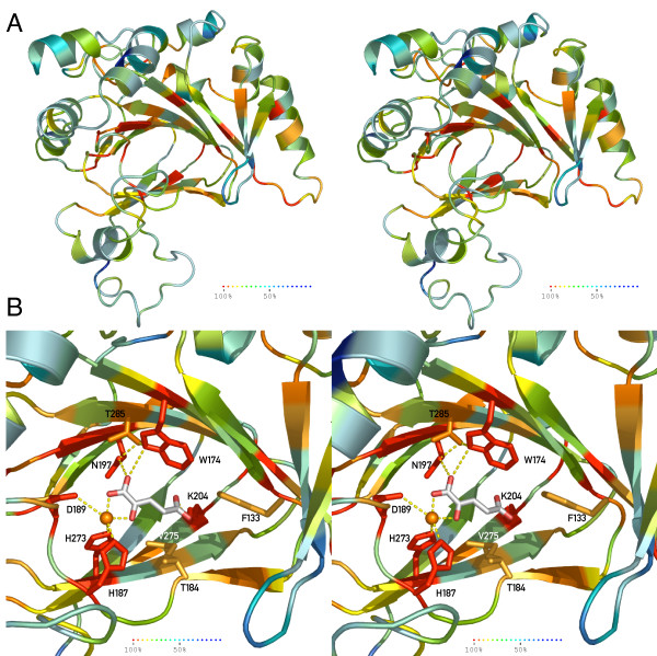
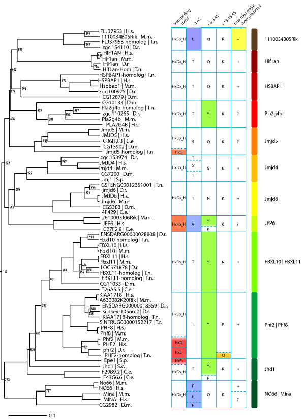
Results: We describe here a semiautomated computational pipeline to identify and annotate JmjC domain containing proteins. Using a sequence segment N-terminal of the Jmjd6 JmjC domain as query for a reciprocal BLAST search, we identified homologous sequences in 62 species across all major phyla. Retrieved Jmjd6 sequences were used to phylogenetically analyse corresponding loci and their genomic neighbourhood. This analysis let to the identification and characterisation of a bi-directional transcriptional unit compromising the Jmjd6 and 1110005A03Rik genes and to the recognition of a new, before overseen Jmjd6 exon in mammals. Using expression studies, two novel Jmjd6 splice variants were identified and validated in vivo. Analysis of the Jmjd6 neighbouring gene 1110005A03Rik revealed an incident deletion of this gene in two out of three earlier reported Jmjd6 knockout mice, which might affect previously described conflicting phenotypes. To determine potentially important residues for Jmjd6 function a structural model of the Jmjd6 protein was calculated based on sequence conservation. This approach identified a conserved double-stranded beta-helix (DSBH) fold and a HxDxnH facial triad as structural motifs. Moreover, our systematic annotation in nine species identified 313 DSBH fold-containing proteins that split into 25 highly conserved subgroups.

Conclusion: We give further evidence that Jmjd6 most likely has a function as a nonheme-Fe(II)-2-oxoglutarate-dependent dioxygenase as previously suggested. Further, we provide novel insights into the evolution of Jmjd6 and other related members of the superfamily of JmjC domain containing proteins. Finally, we discuss possibilities of the involvement of Jmjd6 and 1110005A03Rik in an antagonistic biochemical pathway.


Figures

Figure 1
Figure 1
Computational pipeline flow used for the retrieval and analysis of DSBH/JmjC-domain containing proteins. It contains three parallel procedures. Indicated in yellow colour are searches and analysis approaches based on DNA sequences, data processing approaches based on protein sequences are coloured green, and phylogenomic analysis methods based on DSBH protein domains are shown in blue. Initial queries are depicted as circles, databases as ovals, candidate gene and protein lists as cut-off boxes, and methods/analysis tools as normal boxes, respectively. Barrels indicate Perl script based analysis of retrieved results. Candidate lists of the individual workflows are shown and the final results of the analysis are depicted in bold. Arrows indicate the flow of information and the connections between different search routines and methods. Sources of queried databases and the analysis software used are given in the respective box elements.
Figure 2
Figure 2
Percent identity plot of the chromosomal regions encompassing the Jmjd6 locus in vertebrate species. The genomic regions of eight vertebrate species identified as putative Jmjd6 orthologous loci were aligned with the mouse Jmjd6 locus and its neighbouring genes using PipMaker. Green, blue and yellow overlays highlight the conservation of the annotated exons/coding sequences of the individual genes found in the reference sequence. The orange overlay indicates conserved sequences of the first exons of the Jmjd6 and 1110005A03Rik genes and sequences outside of the coding elements. The red overlay marks a high-scoring segment in all analysed mammalian species supporting the presence of at least one additional exon (No. 5) that is not included in the current Ensembl gene annotation (build 44). Transcriptional orientations of the genes, their exons, UTRs, repetitive elements, and CpG islands are in the symbol key at the figure bottom.
Figure 3
Figure 3
Identification and characterisation of new splice variants of Jmjd6. (A) Two additional putative exons (green boxes) were identified in addition to the transcript annotated in Ensembl (variant 1) by screening all public EST databases. Alternative splicing using these two exons results in the generation of two additional transcripts of Jmjd6 (variants 2 and 3, respectively). Half arrows and orange lines in the schematic presentation of the transcripts highlight the combination of exons detected by RT-PCR. Numbering of Jmjd6 exons includes the additional identified exon variants 4b and 5. (B) Expression analysis of alternative Jmjd6 transcripts in adult mouse organs and embryonic stages (E) using RT-PCR confirmed the presence of all three transcripts at different mRNA expression levels in the analysed samples. Half arrows in (A) indicate the position of the PCR primers used for the amplification. Blue numbers and arrows in (B) point to PCR fragments amplified from splice variants 1 to 3. Amplification of the housekeeping β-actin gene was used as a loading control. (C) The effects of the different splicing events on the C-termini are shown in the gapped alignment from amino acid 300 onwards. The full length human JMJD6 and murine Jmjd6 proteins are represented in the upper part of the figure. Nearly all novel splice variants identified in the mouse (Mm 2) and in humans (Hs 2–3) are truncated at the C-terminus in comparison to splice variant 1 (Mm 1 and Hs 1) and are predicted to contain no poly-serine stretch ([Ser]n). In contrast, the JmjC domain is not affected (green box). Predicted AT-hook domain, sumoylation recognition site (Sumo), and nuclear export signals (NES) are annotated as depicted at the bottom.
Figure 4
Figure 4
Identification of the Jmjd6 1110005A03Rik bi-directional transcriptional unit. (A) Schematic overview of the 65 bp non-coding and non-overlapping intergenic region between the murine Jmjd6 and 1110005A03Rik (Rik) genes. The conserved sequence element identified using PipMaker in Figure 2 is shown in orange. 5'-untranslated regions (UTRs) are indicated in grey, and first exon coding sequences of the Jmjd6 gene in green and of the 1110005A03Rik gene in blue, respectively. The region indicated by two half arrows was used for promoter activity assays presented in (B). It was amplified and cloned to yield an 840 bp test fragment (B) This putative bi-directional promoter fragment was used in a luciferase reporter gene assay to measure transcriptional activity. Basal promoter activity was found for both transcriptional orientations compared to a promoterless control (pGL3) vector. The 5'-3' orientation of the fragment (1110005A03Rik orientation, Rik) yielded only half of the transcriptional activity as it was measured for the 3'-5' orientation (Jmjd6 orientation, Jmjd6). The values reported for the transfection experiments are means ± standard deviation of three independent triplicate experiments. P value was determined using Student's t-test. *** P < 0.001.
Figure 5
Figure 5
Multiple sequence alignment of homologous Jmjd6 proteins. The sequences of 54 homologous Jmjd6 proteins were used for the calculation of a multiple sequence alignment (ClustalW). The numbering of the residues is according to the mouse protein sequence. Identical residues were shaded in blue and similar residues in magenta. Green shading indicates the predicted catalytic residues of the HxDxnH facial. Sequence insertions in individual species outside of the conserved core regions of the mouse Jmjd6 protein were masked out and the maximum numbers of amino acids in these insertions are indicated [n]. Based on the sequence conservation, a frequency corrected sequence logo was calculated (shown above the alignment) and the mouse sequence conservation was highlighted in colour dots according to their degree of conservation (shown as colour code in percent identity below the alignment). The JmjC domain (light blue bar) and additional identified secondary structure elements (red cylinders: α-helices; yellow and green arrows: β-strands) were annotated to the sequence as indicated at the bottom and the β-strands forming the jelly-roll of the JmjC domain were numbered (I-VIII). Green arrows represent the β-sheet that extends the major sheet outside of the jelly-roll.
Figure 6
Figure 6
Predicted structure of Jmjd6 based on comparative modelling (stereoview). Jmjd6 sequence alignments (Fig. 5) and HHpred were used to model Jmjd6 utilising the PDB structures of Hif1an (1h2k), Putative asparaginyl hydroxylase – 2636534 (1vrb) and Jmjd2a (2gp5), respectively. (A) Stereoview of the predicted structure of Jmjd6 presented as a ribbon model. The conserved eight-stranded DSBH core found in all Fe(II) and 2-oxoglutarate (2OG)-dependent dioxygenases is coloured in blue. Additional β-strands attached to the major β-sheet are shown in orange. Helices are depicted in green. (B) View of the predicted active site of the Jmjd6-Fe(II)-2OG complex showing coordination of Fe(II) to 2OG, His187, Asp189, and His273. 2OG also ligates to Trp174, Asn197 and Lys204 with Thr285 stabilising Asn197 and Trp174. Additional important residues for putative interactions are shown in cyan and include hydrophobic interactions from Phe133 and Val275 as well as Thr184, which is involved in 2OG-binding in Hif1an. Interacting residues along with the 2OG co-substrate are shown as stick presentations, putative H-bond interactions as dotted lines. Fe(II) is depicted as an orange ball.
Figure 7
Figure 7
Evolutional conservation of critical residues in Jmjd6 (stereoview). (A) Ribbon presentation of the predicted Jmjd6 structure is coloured based on the sequence homology observed in 54 Jmjd6 homologous proteins (Fig. 5). (B) Annotation of the sequence identity onto the catalytic domain demonstrated the conservation of the critical residues within the active site of the Jmjd6-Fe(II)-2OG complex. Residues were shaded according to the heat map with dark red representing total conservation of residues (100% sequence identity across 54 analysed species), and blue colour representing 50% sequence identity.
Figure 8
Figure 8
Phylogenetic relationship of Jmjd6 to other JmjC domain containing proteins. Detailed section of the phylogenetic tree of 313 JmjC proteins that is presented [in additional file 6]. Shown are 12 out of 25 identified DSBH fold containing protein subgroups as indicated with protein family names on the right side. Scale bar represents the relative phylogenetic distance as determined with PHYLIP. Bootstrap values are shown for values <1000. The first column of the table shows the residues potentially involved in iron binding. The second, third and fourth columns show potentially important residues within the -3, +6–8 and +13–15 regions in respect to the Hx(D/E/H) motif, respectively. Residues discussed in the text are highlighted in red, orange, blue, green and yellow. The fifth column shows the predicted ability to build an extended major sheet with three antiparallel β-strands as determined by Jpred. A "+" indicates the presence of three beta strands, a "?" two beta-strands and a "-" no predicted beta-strands within a 50 aa window starting 80 aa before the first β-strand from the JmjC jelly-roll. H.s. = Homo sapiens, M.m. = Mus musculus, D.r. = Danio rerio, T.n. = Tetraodon nigroviridis, T.r. = Takifugu rubripes, D.m. = Drosophila melanogaster, C.e. = Caenorhabditis elegans, S.c. = Saccharomyces cerevisiae, S.p. = Schizosaccharomyces pombe.

References

    1. Takeuchi T, Yamazaki Y, Katoh-Fukui Y, Tsuchiya R, Kondo S, Motoyama J, Higashinakagawa T. Gene trap capture of a novel mouse gene, jumonji, required for neural tube formation. Genes Dev. 1995;9:1211–1222. doi: 10.1101/gad.9.10.1211. - DOI - PubMed
    1. Balciunas D, Ronne H. Evidence of domain swapping within the jumonji family of transcription factors. Trends Biochem Sci. 2000;25:274–276. doi: 10.1016/S0968-0004(00)01593-0. - DOI - PubMed
    1. Clissold PM, Ponting CP. JmjC: cupin metalloenzyme-like domains in jumonji, hairless and phospholipase A2beta. Trends Biochem Sci. 2001;26:7–9. doi: 10.1016/S0968-0004(00)01700-X. - DOI - PubMed
    1. Klose RJ, Kallin EM, Zhang Y. JmjC-domain-containing proteins and histone demethylation. Nat Rev Genet. 2006;7:715–727. doi: 10.1038/nrg1945. - DOI - PubMed
    1. Takeuchi T, Watanabe Y, Takano-Shimizu T, Kondo S. Roles of jumonji and jumonji family genes in chromatin regulation and development. Dev Dyn. 2006;235:2449–2459. doi: 10.1002/dvdy.20851. - DOI - PubMed

Publication types

MeSH terms

Substances