. 2008 Jun 19:5:50.
doi: 10.1186/1742-4690-5-50.

Reconstitution of the myeloid and lymphoid compartments after the transplantation of autologous and genetically modified CD34+ bone marrow cells, following gamma irradiation in cynomolgus macaques

Affiliations

Reconstitution of the myeloid and lymphoid compartments after the transplantation of autologous and genetically modified CD34+ bone marrow cells, following gamma irradiation in cynomolgus macaques

Sonia Derdouch et al. Retrovirology. .

Abstract

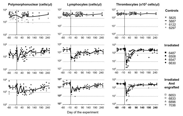
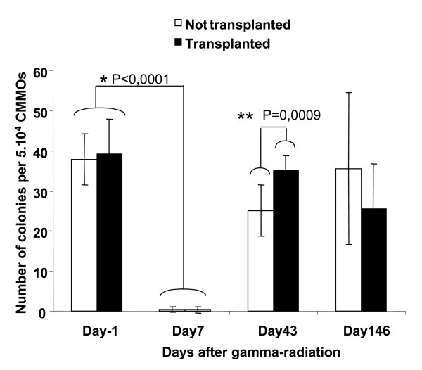
Background: Prolonged, altered hematopoietic reconstitution is commonly observed in patients undergoing myeloablative conditioning and bone marrow and/or mobilized peripheral blood-derived stem cell transplantation. We studied the reconstitution of myeloid and lymphoid compartments after the transplantation of autologous CD34+ bone marrow cells following gamma irradiation in cynomolgus macaques.

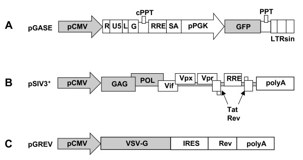
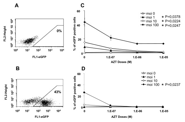
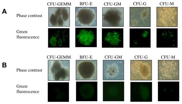
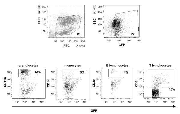
Results: The bone marrow cells were first transduced ex vivo with a lentiviral vector encoding eGFP, with a mean efficiency of 72% +/- 4%. The vector used was derived from the simian immunodeficiency lentivirus SIVmac251, VSV-g pseudotyped and encoded eGFP under the control of the phosphoglycerate kinase promoter. After myeloid differentiation, GFP was detected in colony-forming cells (37% +/- 10%). A previous study showed that transduction rates did not differ significantly between colony-forming cells and immature cells capable of initiating long-term cultures, indicating that progenitor cells and highly immature hematopoietic cells were transduced with similar efficiency. Blood cells producingeGFP were detected as early as three days after transplantation, and eGFP-producing granulocyte and mononuclear cells persisted for more than one year in the periphery.

Conclusion: The transplantation of CD34+ bone marrow cells had beneficial effects for the ex vivo proliferation and differentiation of hematopoietic progenitors, favoring reconstitution of the T- and B-lymphocyte, thrombocyte and red blood cell compartments.


Figures

Figure 1
Figure 1
Schematic representation of SIV-derived SIN vector, helper construct and VSV-g encoding plasmid. An SIVmac251-derived vector was produced by cotransfecting 293T cells with three plasmids: A. a plasmid pGASE containing the eGFP gene under control of the PGK promoter; B. a plasmid pSIV3+ containing viral genes; C. a plasmid pGREV containing the VSV envelope gene. Cis genetic elements are symbolized with white boxes, whereas promoters and genes are depicted by shadowed boxes. pCMV, early cytomegalovirus promoter; pPGK, mouse phosphoglycerate kinase-1 promoter; RRE, REV-responsible element; SA, SIV Rev/Tat splice acceptor; cPPT and PPT, central and 3' polypurine tracks, respectively; GFP, the gene encoding the enhanced green fluorescent protein; LTRsin, partially U3 deleted 3'LTR; LG, leader and a 5' GAG region.
Figure 2
Figure 2
Efficiency of transduction of cynomologus macaque primitive hematopoietic cells with SIV-based lentiviral vectors. A: Non transduced cells were used as a control for each animal. B: Transduction of bone marrow progenitor cells with an SIV-based vector. CD34+ cells were cultured in the presence of cytokines (see materials and methods) and exposed to vector particles at an MOI of 100 for 24 hours before FACS analysis for eGFP production. C: CD34+ cells were cultured overnight in a proliferation medium supplemented with various concentrations of AZT (100 nM, 1 mM, 10 mM). Cells were then washed twice and transduced with various multiplicities of infection (MOI) of the lentiviral vector (0, 1, 10, 100). After 24 hours of coculture with lentiviral vector, some of the CD34+ cells were used to evaluate the rate of transduction of undifferentiated CD34+ cells (C); * indicate statistically significant differences (Kruskal Wallis test) between cultures with and without AZT treatment for MOI = 1 (p = 0,0378), MOI = 10 (p = 0,0224) and MOI = 100 (p = 0,0247). Some of the cells were cultured for 14 days, to allow the myeloid differentiation of CFC. Cells were then resuspended, washed and fixed for three days. They were analyzed by flow cytometry, to evaluate the percentage of eGFP-positive cells and determine the rate of transduction (D); * indicates a statistically significant difference (p = 0,0237(Kruskal Wallis test)) between cultures with and without AZT treatment for MOI = 100. The results shown are the mean values for the three monkeys, each studied in triplicate.
Figure 3
Figure 3
Fluorescence microscopy after myeloid differentiation of CFC (×100). Freshly isolated CD34+ cells were transduced or not with the lentiviral vector (24 hours of culture with lentiviral vector at MOI = 100). Cells were then cultured for 14 days in the presence of cytokines, to allow myeloid differentiation of transduced (A) and not transduced (B) CD34+ cells. Abbreviations: CFU-GEMM, Colony-Forming Unit-Granulocytes, Erythroid, Macrophage, Megakaryocyte; BFU-E, Burst-Forming Unit-Erythroid; CFU-GM, Colony-Forming Unit-Granulocytes, Macrophage; CFU-G, Colony-Forming Unit-Granulocytes; CFU-M, Colony-Forming Unit-Macrophage.
Figure 4
Figure 4
Effect of irradiation and transplantation on polymorphonuclear cell, lymphocyte and thrombocyte counts. All animals were followed during the weeks preceding the study, and for more than 240 days after the irradiation. We carried out hematological analysis including blood cell counts with an automated hemocytometer (Coulter Corporation, Miami, USA).
Figure 5
Figure 5
Recovery of bone marrow clonogenic activity. Bone marrow-derived colony-forming units following sublethal irradiation of cynomolgus monkeys transplanted (black bars) or not transplanted with CD34+ cells (open bars). Mean ± SD of CFC number (triplicate). The results of statistical test are indicates; * indicates a statistically significant difference (p < 0,0001 (Wilcoxon test)) between day 0 and day 7 for the both group; ** indicates a statistically significant difference (p = 0,0009 (Mann & Whitney test)) at day 43 between animals undergoing transplantation and those that did not undergo transplantation.
Figure 6
Figure 6
Flow cytometry analysis of hematopoiesis reconstitution. Animal transplanted with autologous CD34+ bone marrow cells transduced with an SIV-based vector. eGFP-positive cells present in P1 and P2 were analyzed by immuno-staining to identify the subpopulations of eGFP-positive cells in peripheral blood. CD20-PerCP-Cy5, CD14-PE, CD11b-APC and CD3-APC staining were used to identify the B-lymphocyte, monocyte, granulocyte and T-lymphocyte subpopulations.

References

    1. Miller DG, Adam MA, Miller AD. Gene transfer by retrovirus vectors occurs only in cells that are actively replicating at the time of infection. Mol Cell Biol. 1990;10:4239–4242. - PMC - PubMed
    1. Jones RJ, Wagner JE, Celano P, Zicha MS, Sharkis SJ. Separation of pluripotent haematopoietic stem cells from spleen colony-forming cells. Nature. 1990;347:188–189. doi: 10.1038/347188a0. - DOI - PubMed
    1. Gothot A, Loo JC van der, Clapp DW, Srour EF. Cell cycle-related changes in repopulating capacity of human mobilized peripheral blood CD34(+) cells in non-obese diabetic/severe combined immune-deficient mice. Blood. 1998;92:2641–2649. - PubMed
    1. Hao QL, Thiemann FT, Petersen D, Smogorzewska EM, Crooks GM. Extended long-term culture reveals a highly quiescent and primitive human hematopoietic progenitor population. Blood. 1996;88:3306–3313. - PubMed
    1. Ploemacher RE, Sluijs JP van der, Voerman JS, Brons NH. An in vitro limiting-dilution assay of long-term repopulating hematopoietic stem cells in the mouse. Blood. 1989;74:2755–2763. - PubMed

Publication types