

Retinal ganglion cells in diabetes
- PMID: 18565995
- PMCID: PMC2614025
- DOI: 10.1113/jphysiol.2008.156695
Retinal ganglion cells in diabetes
Abstract
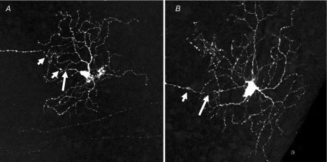
Diabetic retinopathy has long been recognized as a vascular disease that develops in most patients, and it was believed that the visual dysfunction that develops in some diabetics was due to the vascular lesions used to characterize the disease. It is becoming increasingly clear that neuronal cells of the retina also are affected by diabetes, resulting in dysfunction and even degeneration of some neuronal cells. Retinal ganglion cells (RGCs) are the best studied of the retinal neurons with respect to the effect of diabetes. Although investigations are providing new information about RGCs in diabetes, including therapies to inhibit the neurodegeneration, critical information about the function, anatomy and response properties of these cells is yet needed to understand the relationship between RGC changes and visual dysfunction in diabetes.
Figures
References
-
- Abu El-Asrar AM, Dralands L, Missotten L, Geboes K. Expression of antiapoptotic and proapoptotic molecules in diabetic retinas. Eye. 2007;21:238–245. - PubMed
-
- Abu-El-Asrar AM, Dralands L, Missotten L, Al-Jadaan IA, Geboes K. Expression of apoptosis markers in the retinas of human subjects with diabetes. Invest Ophthalmol Vis Sci. 2004;45:2760–2766. - PubMed
-
- Aizu Y, Oyanagi K, Hu J, Nakagawa H. Degeneration of retinal neuronal processes and pigment epithelium in the early stage of the streptozotocin-diabetic rats. Neuropathology. 2002;22:161–170. - PubMed
-
- Ali TK, Matragoon S, Pillai BA, Liou GI, El-Remessy AB. Peroxynitrite mediates retinal neurodegeneration by inhibiting nerve growth factor survival signaling in experimental and human diabetes. Diabetes. 2008;57:889–898. - PubMed
-
- Ambati J, Chalam KV, Chawla DK, D'Angio CT, Guillet EG, Rose SJ, Vanderlinde RE, Ambati BK. Elevated gamma-aminobutyric acid, glutamate, and vascular endothelial growth factor levels in the vitreous of patients with proliferative diabetic retinopathy. Arch Ophthalmol. 1997;115:1161–1166. - PubMed
Publication types
MeSH terms
Grants and funding
LinkOut - more resources
Full Text Sources
Other Literature Sources
Medical