

Tissue sodium concentration in myocardial infarction in humans: a quantitative 23Na MR imaging study
- PMID: 18566171
- PMCID: PMC2572767
- DOI: 10.1148/radiol.2481071027
Tissue sodium concentration in myocardial infarction in humans: a quantitative 23Na MR imaging study
Abstract
Purpose: To prospectively determine whether the absolute tissue sodium concentration (TSC) increases in myocardial infarctions (MIs) in humans and whether TSC is related to infarct size, infarct age, ventricular dysfunction, and/or electrophysiologic inducibility of ventricular arrhythmias.
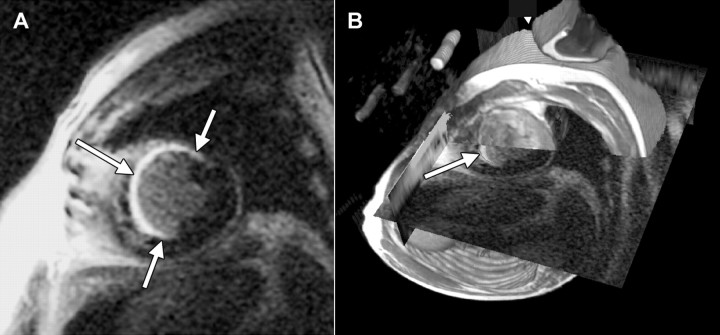
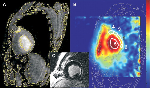
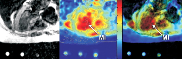
Materials and methods: Delayed contrast material-enhanced 1.5-T hydrogen 1 ((1)H) magnetic resonance (MR) imaging was used to measure the size and location of nonacute MIs in 20 patients (18 men, two women; mean age, 63 years +/- 9 [standard deviation]; age range, 48-82 years) examined at least 90 days after MI. End-systolic and end-diastolic volumes, ejection fraction, and left ventricle (LV) mass were measured with cine MR imaging. The TSC in normal, infarcted, and adjacent myocardial tissue was measured on sodium 23 ((23)Na) MR images coregistered with delayed contrast-enhanced (1)H MR images. Programmed electric stimulation to induce monomorphic ventricular tachycardia (MVT) was used to assess arrhythmic potential, and myocardial TSC was compared between the inducible MVT and noninducible MVT patient groups.
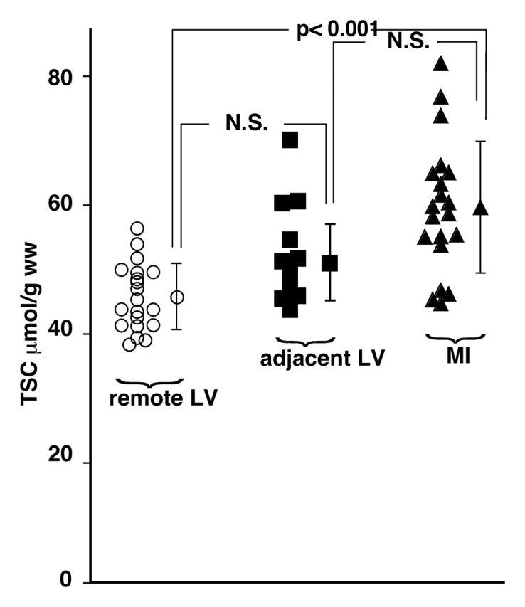
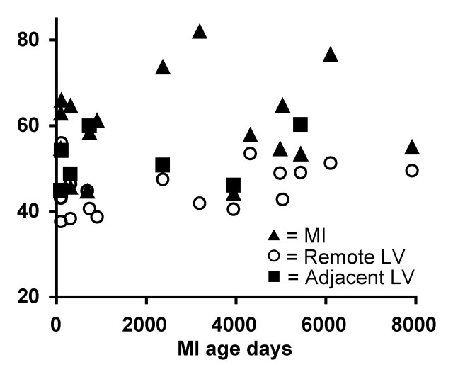
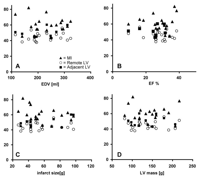
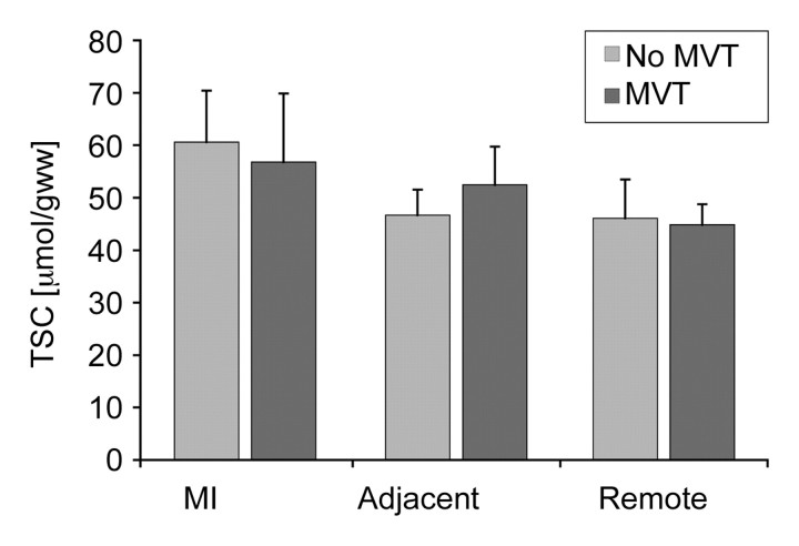
Results: The mean TSC for MIs (59 micromol/g wet weight +/- 10) was 30% higher than that for noninfarcted (remote) LV regions (45 micromol/g wet weight +/- 5, P < .001) and that for healthy control subjects, and TSC did not correlate with infarct age or functional and morphologic indices. The mean TSC for tissue adjacent to the MI (50 micromol/g wet weight +/- 6) was intermediate between that for the MI and that for remote regions. The elevated TSC measured in the MI at (23)Na MR imaging lacked sufficient contrast and spatial resolution for routine visualization of MI. Cardiac TSC did not enable differentiation between patients in whom MVT was inducible and those in whom it was not.
Conclusion: Absolute TSC is measurable with (23)Na MR imaging and is significantly elevated in human MI; however, TSC increase is not related to infarct age, infarct size, or global ventricular function. In regions adjacent to the MI, TSC is slightly increased but not to levels in the MI.
(c) RSNA, 2008.
Figures
References
-
- Zipes DP Genesis of cardiac arrhythmias: electrophysiological considerations. In: Braunwald E, ed. Heart disease: a textbook of cardiovascular medicine. 4th ed. Philadelphia, Pa: Saunders, 1992; 602–627.
-
- Askenasy N, Vivi A, Tassini M, Navon G. Cardiac energetics, cell volumes, sodium fluxes, and membrane permeability: NMR studies of cold ischemia. Am J Physiol 1995;269(3 pt 2):H1056–H1064. - PubMed
-
- Askenasy N, Vivi A, Tassini M, Navon G. The relation between cellular sodium, pH and volumes and the activity of Na/H antiport during hypothermic ischemia: multinuclear NMR studies of rat hearts. J Mol Cell Cardiol 1996;28:589–601. - PubMed
-
- Ouwerkerk R, van Echteld CJ, Staal GE, Rijksen G. Erythrocyte Na+/K+ ATPase activity measured with 23Na NMR. Magn Reson Med 1989;12:164–171. - PubMed
-
- Constantinides CD, Gillen JS, Boada FE, Pomper MG, Bottomley PA. Human skeletal muscle: sodium MR imaging and quantification—potential applications in exercise and disease. Radiology 2000;216:559–568. - PubMed
Publication types
MeSH terms
Substances
Grants and funding
LinkOut - more resources
Full Text Sources
Other Literature Sources
Medical
