. 2008 Sep;26(9):2266-74.
doi: 10.1634/stemcells.2007-1041. Epub 2008 Jun 19.

Human embryonic stem cells have enhanced repair of multiple forms of DNA damage

Affiliations

Human embryonic stem cells have enhanced repair of multiple forms of DNA damage

Scott Maynard et al. Stem Cells. 2008 Sep.

Abstract

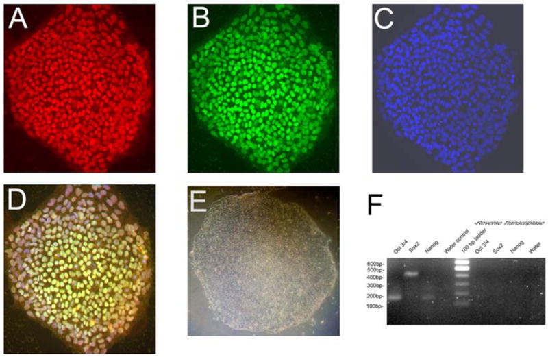
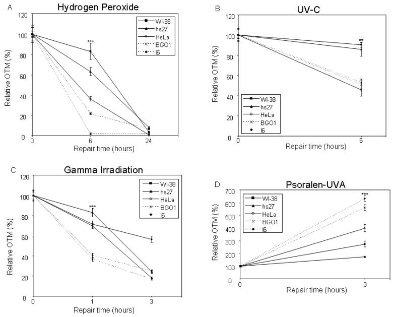
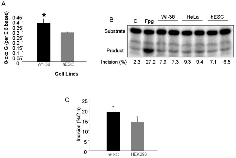
Embryonic stem cells need to maintain genomic integrity so that they can retain the ability to differentiate into multiple cell types without propagating DNA errors. Previous studies have suggested that mechanisms of genome surveillance, including DNA repair, are superior in mouse embryonic stem cells compared with various differentiated murine cells. Using single-cell gel electrophoresis (comet assay) we found that human embryonic stem cells (BG01, I6) have more efficient repair of different types of DNA damage (generated from H2O2, UV-C, ionizing radiation, or psoralen) than human primary fibroblasts (WI-38, hs27) and, with the exception of UV-C damage, HeLa cells. Microarray gene expression analysis showed that mRNA levels of several DNA repair genes are elevated in human embryonic stem cells compared with their differentiated forms (embryoid bodies). These data suggest that genomic maintenance pathways are enhanced in human embryonic stem cells, relative to differentiated human cells.


Figures

Figure 1
Figure 1
Expression of pluripotency markers in undifferentiated hESCs. (A–D) Immunocytochemical analysis of expression of pluripotent markers in hESC line BG01, passage 38: Oct3/4 (A); Sox2 (B); nuclear Hoechst staining (C); and the merged picture (D). (E) Morphology of a typical undifferentiated BG01 hESC colony grown in MEF-CM. (F) RT-PCR on BG01 hESCs with pluripotency markers Oct3/4, Sox2 and Nanog.
Figure 2
Figure 2
Illustration of comet assay design: repair of hydrogen-peroxide induced Fpg-sensitive sites. (A) Olive tail moments (OTM) is the product of the amount of DNA in the tail and the mean distance of migration in the tail. The values for OTM also incorporate subtracted of local background. OTM values were measured as described in Experimental Procedures. (B) Representative assays showing hESC (BG01) and WI-38 cells treated with 100 μM H2O2 and with or without 100 U Fpg, as described in Materials and Methods.
Figure 3
Figure 3
Comet assays for repair of DNA damage induced by hydrogen peroxide, UV-C, IR or psoralen-UVA. The indicated cell types were treated with 100 μM H2O2 (A), 20 J/m2 UV (B), 5 Gy ionizing radiation (C), or 0.2 μg/ml psoralen (30min)/UVA (5 min) (D). Comet assays were then performed as described in Materials and Methods. All 0 h time points were normalized to OTM values of 100%. After the indicated amount of time, H2O2 - and UV-treated cells were treated with 100 U Fpg and 20 U T4 endonuclease V, respectively. Each data point is an average of 50–100 cells calculated as follows, where t represents a given time point after treatment, endo refers to T4 endonuclease, and UT refers to cells not treated with the stress agent indicated: H2O2, (OTM fpgt-OTM no fpgt)-(OTM fpgUT-OTM no fpgUT); UV-C, (OTM endot-OTM no endot)-(OTM endoUT-OTM no endoUT); IR and Psoralen-UVA, OTMt-OTMUT. Data are represented as mean +/− 2xSEM of 50–100 cells. Triple asterisks indicate p<0.01 in hESCs relative to all three other cells lines. Double asterisks indicate p<0.01 in hESCs relative to only the two fibroblasts cells lines (WI-38 and hs27).
Figure 4
Figure 4
Quantification of 8-oxoG and OGG1 incision activity in hESCs. (A) DNA was isolated from hESCs and WI-38, digested with nuclease P1 and alkaline phosphatase, and analyzed by HPLC as described in Experimental Procedures. The value for 8-oxoG per 106 bases is shown. The asterisk indicates significant difference (P=0.02) from the hESC level (B) OGG1 incision assay was performed in duplicate using the indicated cells. C= negative control (no enzyme); Fpg= positive control. Percent incision was calculated as the amount of radioactivity in the product relative to total radioactivity per assay. Background correction was performed using no-enzyme control. (C) Incision assay was performed in triplicate using extracts from hESCs and HEK293. The average incision value +/− SEM was calculated.
Figure 5
Figure 5
Graph of BER gene expression in hESCs relative to embryoid bodies and fibroblasts. Expression levels of repair genes important in BER (A), NER (B), DSB repair (C) and ICL repair (D) are shown. hESC= average expression level of all hESC lines: three hESC lines (BG01, BG02, BG03) and a pool of three other hESC lines (H1, H7, and H9). hEB= average expression level of all human embryoid body (hEB) cell lines: three hEB lines (differentiated from BG01, BG02 and BG03) and a pool of hEB lines differentiated from hESCs H1, H7, and H9. Data are represented as mean +/− SEM.
Figure 6
Figure 6
Western blot analysis of extracts from hESC, WI-38 and HeLa cells. Western blot of hESCs and WI-38 cells treated with 100 μM H2O2 for 30 min. Cells were allowed to recover for the indicated amount of time before preparation of cell extract. The blot were probed with antibody to OGG1, APE-1 and actin (loading control) (A) and band intensities quantitated relative actin (B). Since actin intensity was higher in the hESC line we compared only the change in band intensities.

References

    1. Bohr VA. Repair of oxidative DNA damage in nuclear and mitochondrial DNA, and some changes with aging in mammalian cells. Free Radic Biol Med. 2002;32:804–812. - PubMed
    1. Houtgraaf JH, Versmissen J, van der Giessen WJ. A concise review of DNA damage checkpoints and repair in mammalian cells. Cardiovasc Revasc Med. 2006;7:165–172. - PubMed
    1. Peterson CL, Cote J. Cellular machineries for chromosomal DNA repair. Genes Dev. 2004;18:602–616. - PubMed
    1. Sancar A, Lindsey-Boltz LA, Unsal-Kacmaz K, Linn S. Molecular mechanisms of mammalian DNA repair and the DNA damage checkpoints. Annual Review of Biochemistry. 2004;73:39–85. - PubMed
    1. Andressoo JO, Hoeijmakers JH, Mitchell JR. Nucleotide excision repair disorders and the balance between cancer and aging. Cell Cycle. 2006;5:2886–2888. - PubMed

Publication types