

Roles of heat shock proteins and gamma delta T cells in inflammation
- PMID: 18566334
- PMCID: PMC2574523
- DOI: 10.1165/rcmb.2008-0090TR
Roles of heat shock proteins and gamma delta T cells in inflammation
Abstract
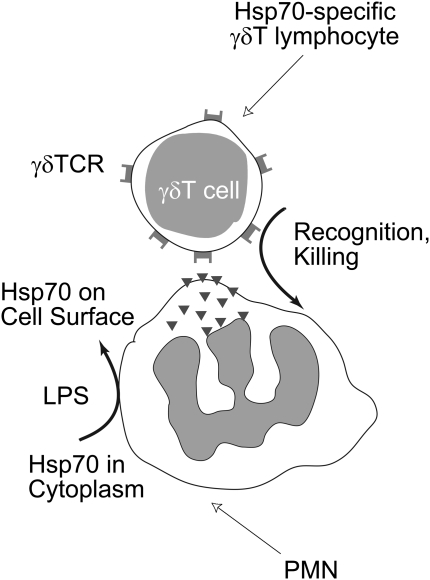
Elimination of activated inflammatory cells that infiltrate and damage host organs can reduce morbidity and mortality. A better understanding of the mechanisms by which these processes occur may lead to new approaches to prevent tissue damage. The lungs, gastrointestinal tract, and skin are particularly prone to infection and collateral damage by inflammatory cells. Specialized lymphocytes protect these organs from collateral tissue damage by eliminating neutrophils and macrophages from inflamed tissues. These lymphocytes recognize signals produced by inflammatory cells. One such signal is heat shock protein (Hsp) expressed on the cell surface of inflamed phagocytes. Mammalian Hsp molecules closely resemble their microbial equivalents, and therefore phagocytes decorated with these molecules are recognized as target cells. T lymphocytes bearing the gammadelta T cell receptor (TCR) elicit cytotoxic activity toward macrophages and neutrophils that express Hsp60 and Hsp70, respectively, protecting host organs from collateral tissue damage by phagocytes.
Figures
References
-
- Rangel-Frausto MS, Pittet D, Costigan M, Hwang T, Davis CS, Wenzel RP. The natural history of the systemic inflammatory response syndrome (SIRS): a prospective study. JAMA 1995;273:117–123. - PubMed
-
- Downey GP, Dong Q, Kruger J, Dedhar S, Cherapanov V. Regulation of neutrophil activation in acute lung injury. Chest 1999;116:46S–54S. - PubMed
-
- Jaeschke H, Smith CW. Mechanisms of neutrophil-induced parenchymal cell injury. J Leukoc Biol 1997;61:647–653. - PubMed
-
- Windsor AC, Mullen PG, Fowler AA, Sugerman HJ. Role of the neutrophil in adult respiratory distress syndrome. Br J Surg 1993;80:10–17. - PubMed
Publication types
MeSH terms
Substances
Grants and funding
LinkOut - more resources
Full Text Sources
Research Materials
Miscellaneous
