. 2008 Jul 1;181(1):73-80.
doi: 10.4049/jimmunol.181.1.73.

Fetal exposure to high-avidity TCR ligand enhances expansion of peripheral T regulatory cells

Affiliations

Fetal exposure to high-avidity TCR ligand enhances expansion of peripheral T regulatory cells

Ping Yu et al. J Immunol. .

Abstract

Lately, it has become clear that regulatory T cells (Tregs) play a major role in the maintenance of peripheral tolerance and control of autoimmunity. Despite these critical functions, the process underlying the development of Tregs remains largely undefined. Herein, altered peptide ligand (APL) variants derived from the proteolipid protein-1 (PLP1) epitope were expressed on immunoglobulins (Igs) and the resulting Ig-APLs were used to deliver the APLs from mother to fetus through the maternal placenta to influence thymic T cell selection. This delivery system was then adapted to the SJL/J mouse, a strain that expresses only the DM20 form of PLP, which lacks the dominant PLP1 epitope in the thymus during fetal and neonatal development. This model, which restores thymic T cell selection for PLP1, was then used to determine whether affinity plays a role in the development of Tregs. The findings show that fetal exposure to low-affinity peptide ligand was unable to drive development of Tregs while variants with higher affinity to the TCR resulted in significant seeding of the periphery with mature, naive Tregs. Thus, contrary to pathogenic T cells, Tregs require avid TCR-ligand interaction to undergo thymic development and maturation.


Figures

Figure 1
Figure 1
Expression of altered peptides on Igs preserves functional avidity. Splenocytes (5 × 10⁵ per well) from 5B6 TCR transgenic mice containing both PLP1-specific T cells and APCs were incubated with graded amounts of Ig chimeras for 3 days. Subsequently, [3H]-thymidine 1 μCi/well was added and the culture continued for an additional 14.5 hours. The cells were then harvested and the incorporated [3H]-thymidine was counted on a Trilux 1450 Microbeta counter. Each point represents the mean of triplicate wells. The results are representative of three independent experiments.
Figure 2
Figure 2
Aggregated Ig-PLP1 is more effective than Ig-PLP-Y or Ig-PLP-LR for expansion of PLP1-specific Tregs. Naïve PLP1-specific 5B6 TCR Tg CD4+ T cells were injected into adult SJL/J mice (5 × 10⁶ cells/mouse) i.v. through the tail vein and the recipients were given an i.p. injection of 300 μg of agg Ig chimeras in saline on days 4, 8, and 12 post transfer. Ten days later, the splenic CD4+ T cells were isolated by positive selection on anti-CD4 Ab coupled to microbeads (Miltenyi Biotec). The CD4 T cells (2 × 10⁶ cells/sample) were analyzed for cell surface expression of TCRVβ6 (left panel), or coexpression of CD25 (middle panel), or CD25 and FoxP3 (right panel) by flow cytometry. The number in the box indicates the percentage of cells expressing the corresponding marker among total CD4 cells. The results are representative of five different experiments.
Figure 3
Figure 3
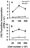
Ig-PLP1 transfers from mother to fetus and drives peptide presentation by fetal thymic and splenic APCs. Pregnant SJL/J female mice were given 300 μg of Ig-PLP1 or control Ig-W on day 19 of gestation, and graded numbers of thymic (A) and splenic (B) cells of the offspring born on day 21 were irradiated and assayed for activation of PLP1-specific TCR transgenic 5B6 CD4+ T cells (5 × 10⁴ cell per well) without adding exogenous antigens. Proliferation was measured by [3H]-Thymidine incorporation. Each point represents the mean ± SD of triplicates.
Figure 4
Figure 4
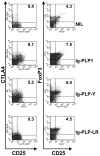
Fetal exposure to high avidity chimeras magnify Treg generation. Pregnant female SJL/J mice were given Ig-PLP1, Ig-PLP-Y or Ig-PLP-LR on day 16, 17 and 18 of gestation and the offspring born on day 21 were allowed to grow into adults. At 6 weeks of age the offspring were given three injections of agg Ig-PLP1 (300 μg/ injection) four days apart and 10 days later the splenic CD4+ T cells were purified and stained for surface CD25 and CTLA4, as well as intracellular FoxP3 as described in Figure 3. One group of mice that had not been exposed to any antigen during fetal life (NIL), but received agg Ig-PLP1 for Tregs expansion was included for control purposes. Cells were gated on live cells and analyzed for CD25, CTLA4 and FoxP3 expression. The numbers in the upper right corner indicate the percentage of cells expressing the specific marker among total gated CD4+ T cells. The results are representative of three different experiments.
Figure 5
Figure 5
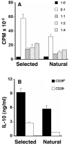
Ig-PLP-Y-selected CD4+CD25+ T cells suppress proliferation of their CD4+CD25- counterparts. SJL/J offspring that had been fetally exposed to Ig-PLP-Y were given agg Ig-PLP1 at the age of 6 weeks and the expanded CD4+CD25+ T cells were purified and tested for suppression of their CD4+CD25- counterparts (A) and IL-10 production (B). The cells were purified ten days after the last injection of agg Ig-PLP1 and separation was done by MACS using CD4 T cell isolation kit and anti-CD25-microbeads according to Miltenyi Biotec’s instructions. For measurement of suppression CD4+CD25+ (2 × 10⁵ T cells per well) were incubated with graded numbers of CD4+CD25- T cells and 4 × 10⁵ SJL-irradiated (3000 rad) splenocytes as APCs. Soluble anti-CD3 antibody (4μg/ml) was then added and the culture was continued for 48 hours. Subsequently [3H]-thymidine was added (1 μCi/well) and proliferation was measured 14.5 hours later. CD4+CD25+ T cells alone without effectors (1:0 black bars) and CD4+CD25- effectors without Tregs (0:1 open bars) were included to serve as controls. CD4+CD25+ T cells from naïve SJL/J mice that were not subject to fetal selection and expansion regimens (Natural) were included for comparison with cells that were fetally selected with Ig-PLP-Y and expanded with agg Ig-PLP-1. Each bar represents the mean ± SD of triplicates. For measurement of IL-10 (B) selected and natural CD4+CD25+ (2 × 10⁵ cells per well) were incubated on plate-bound anti-CD3 Ab (5 μg/ml) for 48 h. IL-10 production was detected by ELISA. CD4+CD25- T cell counterparts (2 × 10⁵ cells per well) were included to serve as control. Incubation on plate bound hamster IgG was performed as isotype control and no measurable IL-10 was detected. Each bar represents the mean ± SD of triplicates. The results are representative of three experiments.
Figure 6
Figure 6
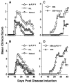
Ig-PLP-Y selected Treg cells ameliorate both passive and active EAE. Six week-old SJL/J mice (A and B) were given natural (diamond) or Ig-PLP-Y selected (triangles) Tregs (1 × 10⁶ cells per mouse) and one day later induced for EAE with PLP1 peptide (A) or central nervous system (CNS) homogenate (B). The mice were monitored for signs of paralysis daily for 40 days. A group of mice that did not receive any T cell transfer (NIL) was included as control. For passive EAE (C and D) RAG-2-/- SJL mice were given i.v. 1 × 10⁶ naive 5B6 TCR Tg CD4+ T cells alone (5B6) to induce the disease (17). To test for suppression the mice received along with 5B6 TCR Tg CD4+ T cells 0.5 × 10⁶ natural (5B6 + Natural) or Ig-PLP-Y-selected (5B6 + Ig-PLP-Y) Tregs. The mice were monitored daily for signs of paralysis for 30 days. Each point represents the mean clinical score of five to six mice.
Figure 7
Figure 7
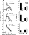
Offspring fetally exposed to high avidity peptides resist EAE induction and produce IL-10 cytokine. Six weeks old SJL/J offspring fetally exposed to Ig-PLP1 (A), Ig-PLP-Y (B) or Ig-PLP-LR (C) were induced for EAE with 100 μg PLP1 peptide in CFA as described in Materials and Methods and monitored daily for signs of paralysis for 30 days. A group of mice that was not exposed to any peptide during fetal life (NIL) was included for control purposes. Each point represents the mean clinical score of five to six mice. Panels (D-F) show IL-10 production by splenic cells from adult SJL/J offspring fetally exposed to (D) Ig-PLP1, (E) Ig-PLP-Y, or (F) Ig-PLP-LR. The mice were immunized with PLP1 peptide in CFA as in A-C except that not pertussis toxin was administered to allow for T cell accumulation in the spleen. Ten days later, the splenic cells (1 × 10⁶ cells/well) were stimulated in vitro with 20 μg/ml PLP1 or the negative control PLP2 peptide and IL-10 secretion was measured by ELISA. A group of mice that were not fetally exposed to any PLP peptide but immunized with PLP1 peptide (NIL) was included for control purpose. Each bar represents the mean ± SD of three to five individually tested mice.
Figure 8
Figure 8
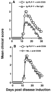
Fetal exposure to Ig-PLP-Y facilitates Tregs selection but does not inactivate pathogenic T cells. SJL/J offspring fetally exposed to Ig-PLP-Y were given 2 injections (1 mg per mouse per injection) of either of anti-CD25 antibody (PC61) (Circles) or Rat- IgG (triangles) on day -4 and -2 before induction of EAE with 100 μg PLP1 peptide in CFA. The animals were then monitored for clinical signs of EAE daily for 30 days. SJL/J offspring that were not fetally exposed to any PLP peptide but received anti-CD25 antibody (diamond) were used as control.

References

    1. Surh CD, Sprent J. T-cell apoptosis detected in situ during positive and negative selection in the thymus. Nature. 1994;372:100–103. - PubMed
    1. Starr TK, Jameson SC, Hogquist KA. Positive and negative selection of T cells. Annu. Rev. Immunol. 2003;21:139–176. - PubMed
    1. Sakaguchi S, Sakaguchi N, Asano M, Itoh M, Toda M. Immunologic self tolerance maintained by T cells expressing IL-2 receptor alpha chains (CD25). Breakdown of a single mechanism of self tolerance causes various autoimmune diseases. J. Immunol. 1995;155:1151–1164. - PubMed
    1. Shevach EM. Regulatory T cells in autoimmunity. Annu. Rev. Immunol. 2000;18:423–449. - PubMed
    1. Itoh M, Takahashi T, Sakaguchi N, Kuniyasu Y, Shimizu J, Otsuka F, Sakaguchi S. Thymus and autoimmunity: production of CD25+CD4+ naturally anergic and suppressive T cells as a key function of the thymus in maintaining immunologic self-tolerance. J. Immunol. 1999;162:5317–5326. - PubMed

Publication types

MeSH terms