. 2008 Jul 1;181(1):235-42.
doi: 10.4049/jimmunol.181.1.235.

Peroxisome proliferator-activated receptor-gamma regulates the expression of alveolar macrophage macrophage colony-stimulating factor

Affiliations

Peroxisome proliferator-activated receptor-gamma regulates the expression of alveolar macrophage macrophage colony-stimulating factor

Tracey L Bonfield et al. J Immunol. .

Abstract

Macrophage CSF (M-CSF) regulates monocyte differentiation, activation, and foam cell formation. We have observed that it is elevated in human pulmonary alveolar proteinosis (PAP) and in the GM-CSF knockout mouse, a murine model for PAP. A potential regulator of M-CSF, peroxisome proliferator-activated receptor-gamma (PPARgamma), is severely deficient in both human PAP and the GM-CSF knockout mouse. To investigate the role of PPARgamma in alveolar macrophage homeostasis, we generated myeloid-specific PPARgamma knockout mice using the Lys-Cre method to knock out the floxed PPARgamma gene. Similar to the GM-CSF-deficient mouse, absence of alveolar macrophage PPARgamma resulted in development of lung pathology resembling PAP in 16-wk-old mice, along with excess M-CSF gene expression and secretion. In ex vivo wild-type alveolar macrophages, we observed that M-CSF itself is capable of inducing foam cell formation similar to that seen in PAP. Overexpression of PPARgamma prevented LPS-stimulated M-CSF production in RAW 264.7 cells, an effect that was abrogated by a specific PPARgamma antagonist, GW9662. Use of proteasome inhibitor, MG-132 or a PPARgamma agonist, pioglitazone, prevented LPS-mediated M-CSF induction. Using chromatin immunoprecipitation, we found that PPARgamma is capable of regulating M-CSF through transrepression of NF-kappaB binding at the promoter. Gel-shift assay experiments confirmed that pioglitazone is capable of blocking NF-kappaB binding. Taken together, these data suggest that M-CSF is an important mediator of alveolar macrophage homeostasis, and that transcriptional control of M-CSF production is regulated by NF-kappaB and PPARgamma.


Figures

FIGURE 1
FIGURE 1
PPARγ knockout alveolar macrophage Pathology resembles the GM-CSF KO model. GM-CSF KO (A), PPARγ KO (B), and C57BL/6 control (C) mice were sacrificed, and the lungs stained with periodic-acid Schiff (×40). The GM-CSF KO model demonstrates scattered foci of intraalveolar PAS-positive material (thick arrows, A) and lipid-engorged alveolar macrophages (thick arrow, ×100 inset). All of the PPARγ KO mice (7 of 7 mice) exhibited similar PAS-positive material in alveolar macrophages (B, ×100 inset). Similar to the GM-CSF KO animals, several of these mice (2 of 7) also developed intraalveolar PAS-positive material (B, thin arrow).
FIGURE 2
FIGURE 2
M-CSF induces alveolar macrophages to take up surfactant. Alveolar macrophages were obtained from C57BL/6 mice and cultured in vitro with 200 ng/ml surfactant obtained from a PAP patient in the presence and absence of human recombinant M-CSF. Figures depict ×40 images, representative of six independent experiments. Both surfactant (p = 0.001 vs controls) and M-CSF (p = 0.008) induced Oil-Red-O-positive cells. Alveolar macrophages incubated with surfactant and M-CSF had a cumulatively greater uptake of Oil-Red-O staining lipids compared with either condition alone (p = 0.03 vs either). Each condition was evaluated for (and counted in 6 frames of at least 200 cells) by blinded numerical designation. Data represent mean percentage of positive Oil-Red-O staining ± SEM (n = 6). Abs to M-CSF neutralized the effect of M-CSF on surfactant uptake to levels comparable to surfactant alone. US, Unstimulated; S, surfactant.
FIGURE 3
FIGURE 3
M-CSF gene expression and protein are elevated in BAL specimens from GM-CSF and PPARγ KO mice. Acellular BAL fluid was assessed for M-CSF protein by ELISA, and BAL cell pellets were evaluated for M-CSF mRNA. Compared with wild-type mice (n = 5), BAL cells from both GMCSF KO (n = 4) and PPARγ KO (n = 4) mice demonstrated increased M-CSF gene expression (p ≤ 0.002 for each comparison, B). Similarly, BAL protein levels were increased in both models when compared with wild-type mice (B, p < 0.002, n = 4 per condition).
FIGURE 4
FIGURE 4
Transfection of PPARγ expression vector into RAW 264.7 cells attenuates LPS-mediated induction of M-CSF. RAW 264.7 cells were transfected with 1 μg PPARγ murine expression vector. We measured the expression of M-CSF after 24 h in the presence and absence of LPS. Transient transfection of the PPARγ expression vector resulted in 10,809 ± 1,578% (mean ± SEM) increased expression of PPARγ mRNA expression compared with the control (β-gal) vector (right bars, n = 3, p ≤ 0.001). Compared with transfected controls (β-gal), LPS-induced M-CSF production was inhibited by 90.1 ± 13.2% with the overexpression of PPARγ expression vector (left bars, p = 0.017, n = 3).
FIGURE 5
FIGURE 5
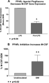
M-CSF secretion is enhanced by PPARγ antagonists and suppressed by PPARγ agonist. RAW cells were cultured in the presence and absence of pioglitazone or GW9662 at baseline or after stimulation with LPS. LPS-stimulated RAW cells treated with pioglitazone expressed 41 ± 6% significantly less M-CSF mRNA (A, p = 0.04, n = 4) and 12 ± 0.7% less M-CSF protein (p = 0.01) compared with the LPS cultures alone. Conversely, baseline RAW cells treated with GW9662 increased their M-CSF gene expression and protein by 532 ± 242% (B, p = 0.02, n = 3) and 11.2 ± 1% (p = 0.03), respectively.
FIGURE 6
FIGURE 6
PPARγ regulates M-CSF through NF-κB. RAW 264.7 cells were stimulated with LPS, which resulted in a 21.6 ± 0.9-fold increase in M-CSF expression. LPS induced M-CSF expression was reduced by 60 ± 26% when the NF-κB was inhibited with the proteasomal blocker. Pioglitazone treatment also decreased LPS-stimulated M-CSF expression, by 68 ± 15% of the LPS control (p < 0.05 vs LPS alone). The combination of both pioglitazone and NF-κB inhibition resulted in 80 ± 25% reduction of M-CSF expression (p < 0.001 vs LPS). These data suggest that PPARγ can regulate M-CSF induction through repression of NF-κB.
FIGURE 7
FIGURE 7
Pioglitazone inhibits LPS-mediated NF-κB activation. Whole-cell extracts were analyzed by EMSA using a 32P-labeled oligonucleotide containing the κB consensus sequences and analyzed on a 4% nondenaturing acrylamide gel. Specificity of the band was confirmed by supershift analysis with p50 and p65 Abs and competition with cold oligonucleotide. Representative image from four independent experiments is shown. Quantification using the StormImager with ImageQuant analysis revealed that pretreatment with pioglitazone before addition of LPS decreased NF-κB binding by 30.6 ± 6.9% (mean (SEM), p < 0.01 by Tukey's test). US, Unstimulated; Pio, pioglitazone.
FIGURE 8
FIGURE 8
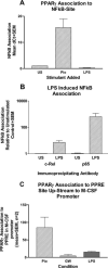
PPARγ inhibits LPS-induced NF-κB association with the M-CSF promoter. ChIP was performed using BALB/c bone marrow-derived macrophages. After treatment with LPS (2 μg/ml), pioglitazone (Pio, 10 μg/ml), or GW9662 (GW, 10 mM), cell lysates were subjected to immunoprecipitation with anti-PPARγ, anti-c-Rel, or anti-p65. Binding to the M-CSF promoter was determined by real-time PCR. LPS stimulation resulted in c-Rel and p65 binding to the NF-κB site upstream of the M-CSF promoter (B). Activation of PPARγ by pioglitazone increased PPARγ association with the NF-κB response element, which was nearly eliminated with NF-κB activation by LPS (A). Pioglitazone also resulted in PPARγ binding to the M-CSF PPRE promoter region (C).
FIGURE 9
FIGURE 9
Model of PPARγ regulation of M-CSF transcription. Data from the ChIP and culture studies were put together to develop a model of a working hypothesis in which PPARγ regulates M-CSF through NF-κB. PPARγ inhibits association of NF-κB proteins with the promoter, an effect that is enhanced in the presence of PPARγ activation. In the presence of NF-κB activation, PPARγ binding to the promoter is down-regulated unless PPARγ is ligand-activated by pioglitazone. In the absence of PPARγ, there is a potential constitutive production of M-CSF through p65/c-Rel binding to NF-κB transcription site.

References

    1. Rotondo D, Davidson J. Prostaglandin and PPAR control of immune cell function. Immunology. 2002;105:20–22. - PMC - PubMed
    1. Berger J, Moller DE. The mechanisms of action of PPARs. Annu. Rev. Med. 2002;53:409–435. - PubMed
    1. Clark RB. The role of PPARs in inflammation and immunity. J. Leukocyte Biol. 2002;71:388–400. - PubMed
    1. Chinetti G, Griglio S, Antonucci M, Torra IP, Delerive P, Majd Z, Fruchart J-C, Chapman J, Najib J, Staels B. Activation of proliferator-activated receptors alpha and gamma induces apoptosis of human monocyte-derived macrophages. J. Biol. Chem. 1998;273:25573–25580. - PubMed
    1. Bonfield TL, Russell D, Burgess S, Malur A, Kavuru MS, Thomassen MJ. Autoantibodies against granulocyte macrophage colony-stimulating factor are diagnostic for pulmonary alveolar proteinosis. Am. J. Respir. Cell Mol. Biol. 2002;27:481–486. - PubMed

MeSH terms