. 2008 Jul 1;181(1):546-56.
doi: 10.4049/jimmunol.181.1.546.

Differential CMV-specific CD8+ effector T cell responses in the lung allograft predominate over the blood during human primary infection

Affiliations

Differential CMV-specific CD8+ effector T cell responses in the lung allograft predominate over the blood during human primary infection

Matthew R Pipeling et al. J Immunol. .

Abstract

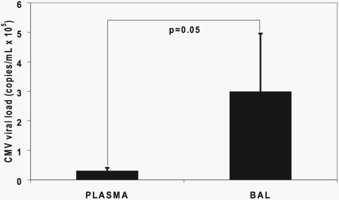
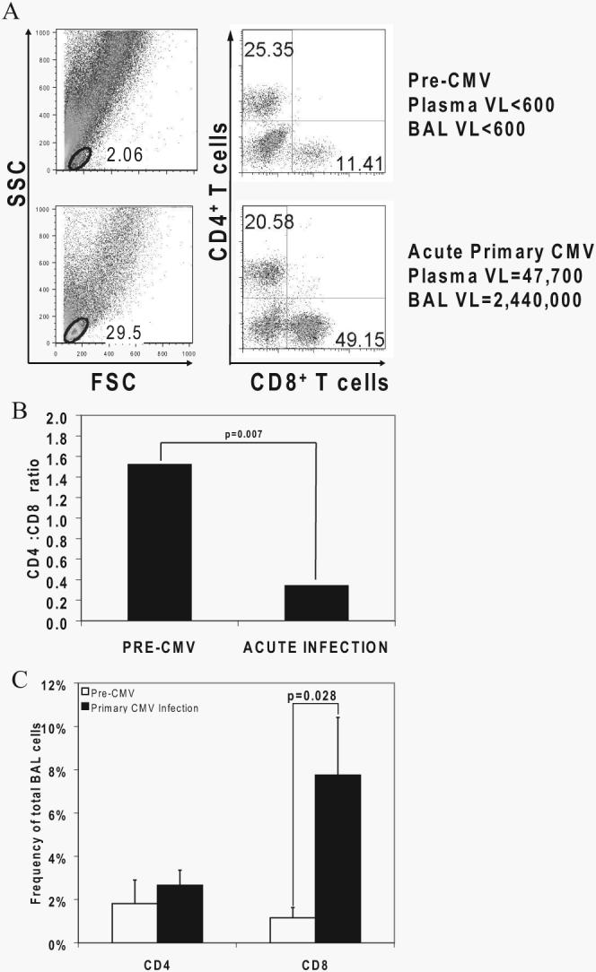
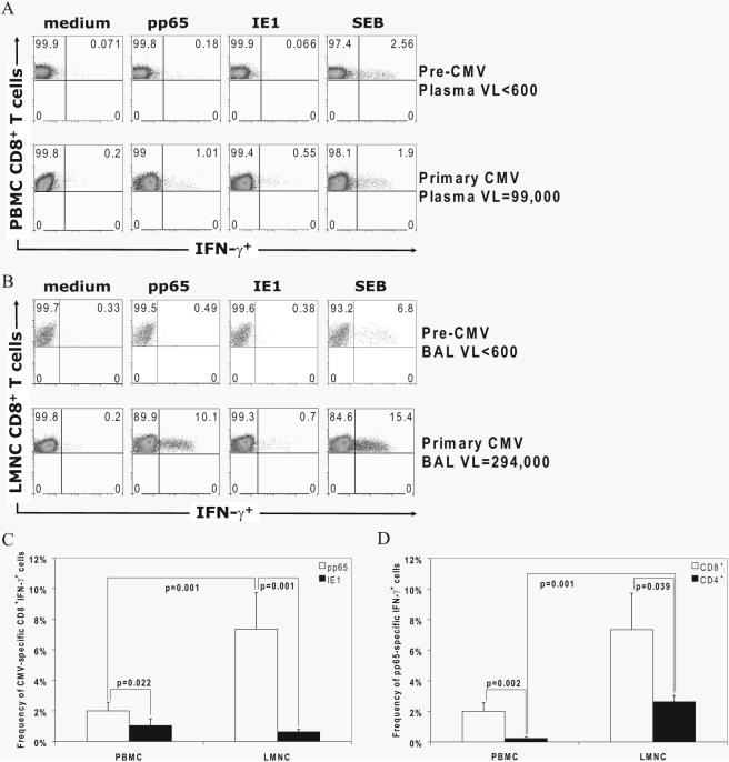
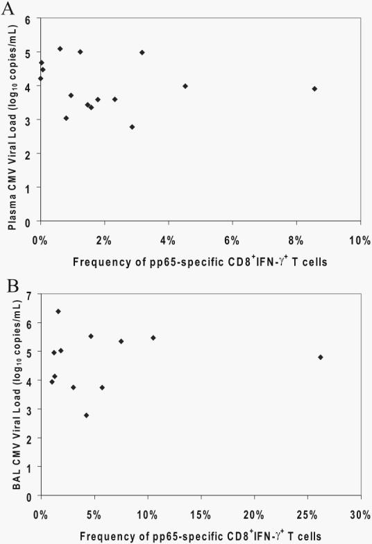
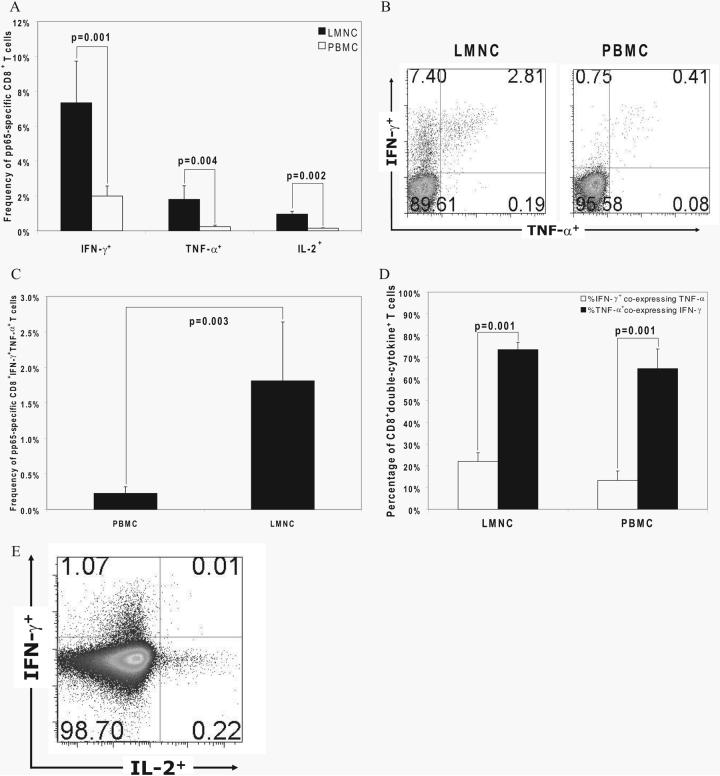
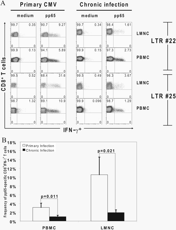
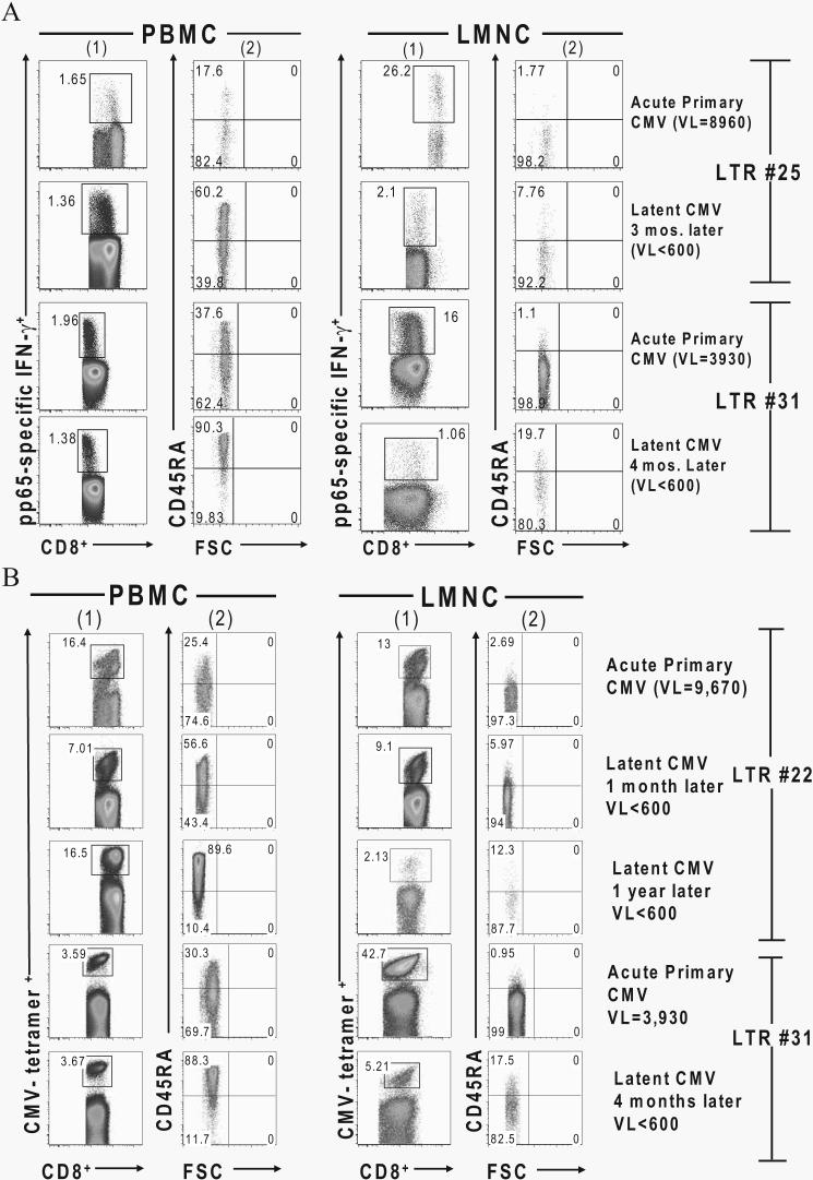
Acquisition of T cell responses during primary CMV infection in lung transplant recipients (LTRs) appear critical for host defense and allograft durability, with increased mortality in donor+/recipient- (D+R-) individuals. In 15 D+R- LTRs studied, acute primary CMV infection was characterized by viremia in the presence or absence of pneumonitis, with viral loads higher in the lung airways/allograft compared with the blood. A striking influx of CD8+ T cells into the lung airways/allograft was observed, with inversion of the CD4+:CD8+ T cell ratio. De novo CMV-specific CD8+ effector frequencies in response to pooled peptides of pp65 were strikingly higher in lung mononuclear cells compared with the PBMC and predominated over IE1-specific responses and CD4+ effector responses in both compartments. The frequencies of pp65-specific cytokine responses were significantly higher in lung mononuclear cells compared with PBMC and demonstrated marked contraction with long-term persistence of effector memory CD8+ T cells in the lung airways following primary infection. CMV-tetramer+CD8+ T cells from PBMC were CD45RA- during viremia and transitioned to CD45RA+ following resolution. In contrast, CMV-specific CD8+ effectors in the lung airways/allograft maintained a CD45RA- phenotype during transition from acute into chronic infection. Together, these data reveal differential CMV-specific CD8+ effector frequencies, immunodominance, and polyfunctional cytokine responses predominating in the lung airways/allograft compared with the blood during acute primary infection. Moreover, we show intercompartmental phenotypic differences in CMV-specific memory responses during the transition to chronic infection.


Figures

FIGURE 1
FIGURE 1
CMV viral loads are increased in the lung allograft compared with the plasma during primary infection. CMV viral loads in the plasma and BAL fluid were determined for the cohort of LTRs by quantitative PCR. Bars represent mean ± SEM viral loads in the respective compartments, and the p value was calculated by Wilcoxon signed-rank test.
FIGURE 2
FIGURE 2
Massive CD8+ T cell expansion occurs in the lung allograft during primary CMV infection with inversion of the CD4+:CD8+ T cell ratio at the time of diagnosis. A, Representative plots of LMNC from LTR no. 29 at pre-CMV and primary CMV time points showing frequencies of CD3+CD4+ and CD3+CD8+ T cells from the lymphoid gates determined by forward scatter (FSC) and side scatter (SSC). VL, Viral load. B, Bars represent geometric means of the CD4+:CD8+ T cell ratio of LMNC from pre-CMV and acute primary CMV infection time points in 10 LTRs, and the p value was calculated by Wilcoxon signed-rank test. C, Bars represent frequencies of CD3+CD4+ and CD3+CD8+ T cells in BAL cells from 10 LTRs at pre-CMV and acute primary CMV infection time points, with p values calculated by Wilcoxon signed-rank test.
FIGURE 3
FIGURE 3
Acquisition of de novo CMV-specific effector T cell responses in the LMNC and PBMC is predominated by pp65-specific CD8+IFN-γ+ effectors in the lung airways/allograft during acute primary CMV infection. A and B, Representative plots of PBMC (A) or LMNC (B) from LTR no. 23 at pre-CMV and acute primary CMV time points are shown. Cells were cultured in medium alone or in the presence of either pp65- or IE1-pooled peptides or the positive control staphylococcal enterotoxin B (SEB) followed by ICS as detailed in Materials and Methods. Quadrant numbers indicate frequencies of populations, with gating on CD3+CD8+ T cells. VL, Viral load. C and D, Frequencies of CD3+CD8+IFN-γ+ pp65-specific and IE1-specific effector responses in LMNC vs PBMC (C) and frequencies of pp65-specific CD3+CD8+IFN-γ+ or CD3+CD4+ IFN-γ+ T cells were determined following in vitro restimulation with pooled pp65 or IE1 peptides. Bars represent mean ± SEM frequency of IFN-γ+ LMNC or PBMC from 15 LTRs, with p values calculated by Wilcoxon signed-rank test.
FIGURE 4
FIGURE 4
CMV viral loads in the plasma and BAL fluid do not correlate with compartmental frequencies of pp65-specific CD8+IFN-γ+ effectors at primary CMV diagnosis. CMV viral loads in the plasma (A) and BAL fluid (B) were determined by quantitative PCR. Frequencies of pp65-specific CD8+ PBMC (A) and LMNC (B) were determined by ICS for IFN-γ. Values for each of 15 or 12 LTRs, respectively, were subjected to scatterplot analyses in the blood (A) and lung (B).
FIGURE 5
FIGURE 5
Hierarchal production of effector cytokines by CD8+ T cells in LMNC and PBMC, with a higher polyfunctional capacity in LMNC. A, Production of IFN-γ, TNF-α, and IL-2 by CD8+ T cells was determined by ICS after in vitro restimulation with pooled pp65 peptides. Bars represent mean ± SEM frequency of cytokine+CD8+ T cells in LMNC and PBMC. B, Representative plot of CD8+ LMNC and PBMC from LTR no. 23 showing frequency of IFN-γ and TNF-α production to pp65. Quadrant numbers indicate frequencies of populations, with gating on CD3+CD8+ T cells. C, Frequencies of CD8+IFN-γ+TNF-α+ LMNC and PBMC from 15 LTRs following in vitro restimulation with pooled pp65 peptides. Bars represent mean ± SEM frequency of CD8+ T cells that coproduce IFN-γ and TNF-α, with p value calculated by Wilcoxon signed-rank test. D, Percentage of CD8+ double-cytokine+ T cells (IFN-γ+ and TNF-α+ detected following gating on IFN-γ+ or TNF-α+ cells in the LMNC or PBMC. E, Representative plot of CD8+ PBMC from LTR no. 34 showing the frequency of IFN-γ and IL-2 production to pp65. Quadrant numbers indicate frequencies of populations with gating on CD3+CD8+ T cells. All p values were determined by Wilcoxon signed-rank test.
FIGURE 6
FIGURE 6
Primary pp65-specific CD8+ effector responses contract in the transition from active to chronic infection but persist long term. A, Representative plots of CD8+IFN-γ+ effector T cell responses in the presence of absence of pooled pp65 peptides by ICS from LMNC and PBMC in LTRs no. 22 and no. 25 during active primary infection compared with latency 1 mo (LTR no. 22) or 3 mo (LTR no. 25) later. Gating is on CD3+CD8+ T cells. B, Contraction of pp65-specific CD8+IFN-γ+ effector T cell frequencies in LMNC and PBMC from nine LTRs during acute primary infection and 3–6 mo postinfection. All p values were determined by Wilcoxon signed-rank test.
FIGURE 7
FIGURE 7
CMV-specific CD8+ T cells transition from a CD45RA phenotype in the PBMC during viremia/acute infection to CD45RA+ during latent infection but remain CD45RA in the lung allograft. A, Representative plots of gated pp65-specific CD8+ IFN-γ+ effector T cells (columns 1) in LMNC and PBMC from LTRs no. 25 and no. 31 during acute primary infection and at latent infection time points following in vitro restimulation with pooled pp65 peptides. CD45RA surface expression by FSC is shown for respective effector gates in columns 2. B, Representative plots of CD8+ CMV tetramer+ T cells in LMNC and PBMC from LTRs no. 22 (both HLA-A2 and HLA-B7 pp65 tetramers) and no. 31 (HLA-A2 pp65 tetramer) at the indicated time points during the transition from acute primary CMV infection into chronic infection. Gating is on CD3+CD8+ tetramer+ T cells. VL, Viral load.

References

    1. Fishman JA, Rubin RH. Infection in organ-transplant recipients. N. Engl. J. Med. 1998;338:1741–1751. - PubMed
    1. Kotloff RM, Ahya VN, Crawford SW. Pulmonary complications of solid organ and hematopoietic stem cell transplantation. Am. J. Respir. Crit. Care Med. 2004;170:22–48. - PubMed
    1. Rubin RH. Cytomegalovirus in solid organ transplantation. Transpl. Infect. Dis. 2001;3(Suppl 2):1–5. - PubMed
    1. Estenne M, Maurer JR, Boehler A, Egan JJ, Frost A, Hertz M, Mallory GB, Snell GI, Yousem S. Bronchiolitis obliterans syndrome 2001: an update of the diagnostic criteria. J. Heart Lung Transplant. 2002;21:297–310. - PubMed
    1. Westall GP, Michaelides A, Williams TJ, Snell GI, Kotsimbos TC. Bronchiolitis obliterans syndrome and early human cytomegalovirus DNAaemia dynamics after lung transplantation. Transplantation. 2003;75:2064–2068. - PubMed

Publication types

MeSH terms