Comparative Study
. 2008 Nov;49(11):5048-54.
doi: 10.1167/iovs.08-1877. Epub 2008 Jun 19.

Quantitative optical coherence tomography findings in various subtypes of neovascular age-related macular degeneration

Affiliations
Comparative Study

Quantitative optical coherence tomography findings in various subtypes of neovascular age-related macular degeneration

Sandra Liakopoulos et al. Invest Ophthalmol Vis Sci. 2008 Nov.

Abstract

Purpose: To compare the volume of various spaces visible on optical coherence tomography (OCT) images in different angiographic lesion subtypes of neovascular age-related macular degeneration (AMD).

Methods: Sixty-six cases of previously untreated, active subfoveal choroidal neovascularization (CNV) associated with AMD were retrospectively collected. CNV lesions were classified as occult with no classic CNV, minimally classic CNV, predominantly classic CNV, or CNV lesions with associated retinal angiomatous proliferation (RAP). Corresponding OCT image sets were analyzed by trained graders using previously validated custom software that allows manual placement of boundaries on OCT B-scans. Spaces delineated by these boundaries included the neurosensory retina, subretinal fluid, subretinal tissue, and pigment epithelial detachments (PEDs). Volume measurements were calculated by the software and compared among groups.

Results: Minimally and predominantly classic CNV membranes demonstrated subretinal tissue on OCT in all cases and appeared to show a significantly greater volume of subretinal tissue than did the occult membranes. Subretinal fluid was present in all the predominantly classic cases. A PED was visible in all the occult CNV cases in our study, demonstrating less retinal thickening and significantly greater PED volumes than minimally and predominantly classic CNV lesions. Lesions associated with RAP showed the highest percentage of cystoid spaces.

Conclusions: OCT and angiography provide complementary information regarding CNV lesions. Quantitative analysis of OCT images allows for an improved understanding of the anatomic characteristics of angiographically defined CNV lesion subtypes.


Figures

Figure 1
Figure 1
OCTOR map demonstrating the volume and thickness of all spaces calculated by the OCTOR software after manual grading of all 6 OCT B-scans.
Figure 2
Figure 2
Fluorescein angiographic (FA) frames [early phase (A), mid phase (B), late phase (C)] of a case with minimally classic CNV. The corresponding OCT B-scan (D, scan direction marked by red arrow in C) demonstrates subretinal fluid, subretinal tissue and a pigment epithelial detachment (PED). Of note, OCT illustrates that the PED (presumably corresponding to Type 1 CNV) continues underneath the subretinal tissue (presumably corresponding to Type 2 CNV), while on FA, only a circular area of stippled hyperfluorescence is visible indicating occult CNV, while the central part of the PED is hidden underneath classic CNV.
Figure 3
Figure 3
Fluorescein angiographic (FA) frames [early phase (A), mid phase (B), late phase (C)] of a case with occult with no classic choroidal neovascularization (CNV). The corresponding optical coherence tomography (OCT) B-Scan (D, scan direction marked by red arrow in C) shows a pigment epithelial detachment and subretinal fluid.
Figure 4
Figure 4
Fluorescein angiographic (FA) frames [early phase (A), mid phase (B), late phase (C)] of a case demonstrating a stereoscopically visible retinal angiomatous proliferation (RAP) connected to occult choroidal neovascularization (CNV). The corresponding optical coherence tomography (OCT) B-Scan (C) shows cystoid retinal edema and subretinal tissue. The RAP lesion itself is not captured by the OCT B-scan.
Figure 5
Figure 5
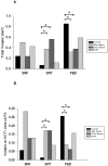
Comparison between angiographic lesion subtypes regarding the total volume of the different spaces visible on Optical Coherence Tomography (OCT) images. Eyes with retinal angiomatous proliferation (RAP) showed the largest total retinal volume, eyes with classic choroidal neovascularization (CNV) components showed the largest volume of subretinal tissue and eyes with occult with no classic CNV showed the largest volumes of pigment epithelial detachments. *Statistically significant difference between groups (p<0.001) A: Total volume values of each space used for analysis B: Total volume of each space devided by area of the total CNV lesion (volume / area ratio) used for analysis
Figure 6
Figure 6
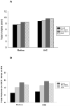
Comparison between angiographic lesion subtypes regarding the total volume of the different spaces visible on Optical Coherence Tomography (OCT) images. There was no statistically significant difference between groups regarding the total volume of the retina or the inner retinal height from the choroid (IHC). Eyes with retinal angiomatous proliferation (RAP) showed the largest total retinal volume, eyes with classic choroidal neovascularization (CNV) components showed the largest volume of subretinal tissue and eyes with occult with no classic CNV showed the largest volumes of pigment epithelial detachments. *Statistically significant difference between groups (p<0.001) A: Total volume values of each space used for analysis B: Total volume of each space devided by area of the total CNV lesion (volume / area ratio) used for analysis

References

    1. Bressler NM, Bressler SB, Congdon NG, Ferris FL, 3rd, Friedman DS, Klein R, et al. Age-Related Eye Disease Study Research Group. Potential public health impact of Age-Related Eye Disease Study results: AREDS report no. 11. Arch Ophthalmol. 2003;121:1621–1624. - PMC - PubMed
    1. Barbazetto I, Burdan A, Bressler NM, et al. Treatment of Age-Related Macular Degeneration with Photodynamic Therapy Study Group. Verteporfin in Photodynamic Therapy Study Group Photodynamic therapy of subfoveal choroidal neovascularization with verteporfin: fluorescein angiographic guidelines for evaluation and treatment--TAP and VIP report No. 2. Arch Ophthalmol. 2003;121:1253–68. - PubMed
    1. Kuhn D, Meunier I, Soubrane G, et al. Imaging of chorioretinal anastomoses in vascularized retinal pigment epithelium detachments. Arch Ophthalmol. 1995;11:1392–8. - PubMed
    1. Brown DM, Kaiser PK, Michels M, et al. ANCHOR Study Group Ranibizumab versus verteporfin for neovascular age-related macular degeneration. N Engl J Med. 2006;355:1432–44. - PubMed
    1. Rosenfeld PJ, Brown DM, Heier JS, et al. MARINA Study Group Ranibizumab for neovascular age-related macular degeneration. N Engl J Med. 2006;355:1419–31. - PubMed

Publication types