. 2008 Jul 23;27(14):1974-84.
doi: 10.1038/emboj.2008.122. Epub 2008 Jun 19.

Stress-protective signalling of prion protein is corrupted by scrapie prions

Affiliations

Stress-protective signalling of prion protein is corrupted by scrapie prions

Angelika S Rambold et al. EMBO J. .

Abstract

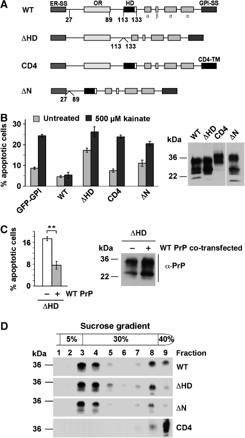
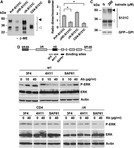
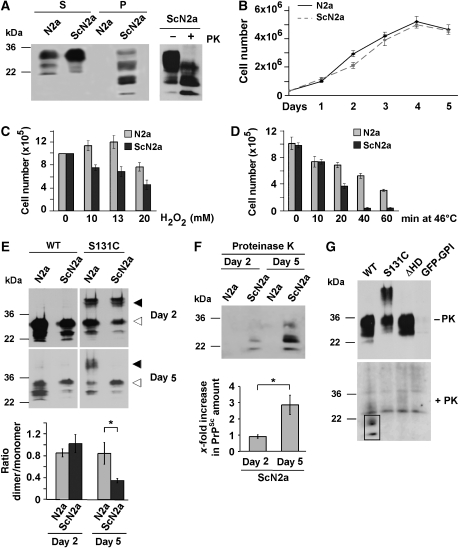
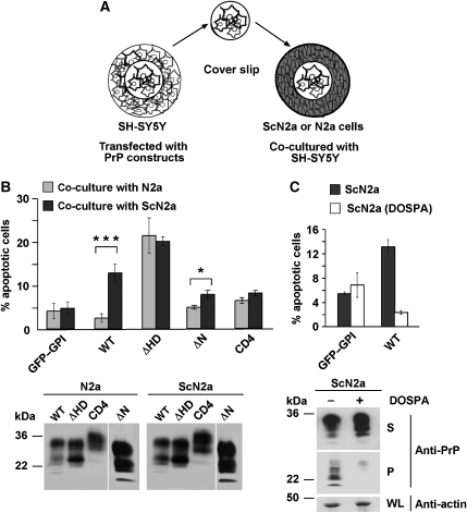
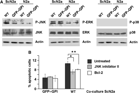
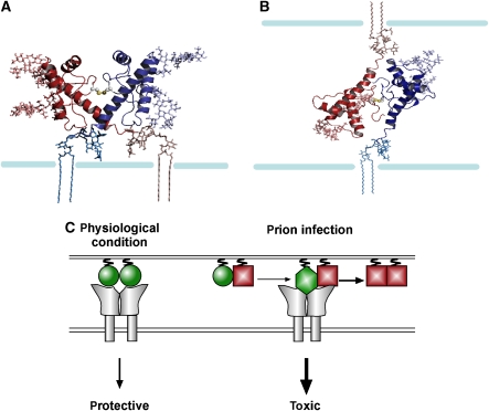
Studies in transgenic mice revealed that neurodegeneration induced by scrapie prion (PrP(Sc)) propagation is dependent on neuronal expression of the cellular prion protein PrP(C). On the other hand, there is evidence that PrP(C) itself has a stress-protective activity. Here, we show that the toxic activity of PrP(Sc) and the protective activity of PrP(C) are interconnected. With a novel co-cultivation assay, we demonstrate that PrP(Sc) can induce apoptotic signalling in PrP(C)-expressing cells. However, cells expressing PrP mutants with an impaired stress-protective activity were resistant to PrP(Sc)-induced toxicity. We also show that the internal hydrophobic domain promotes dimer formation of PrP and that dimerization of PrP is linked to its stress-protective activity. PrP mutants defective in dimer formation did not confer enhanced stress tolerance. Moreover, in chronically scrapie-infected neuroblastoma cells the amount of PrP(C) dimers inversely correlated with the amount of PrP(Sc) and the resistance of the cells to various stress conditions. Our results provide new insight into the mechanism of PrP(C)-mediated neuroprotection and indicate that pathological PrP conformers abuse PrP(C)-dependent pathways for apoptotic signalling.


Figures

Figure 1
Figure 1
The internal hydrophobic domain and the GPI anchor are necessary for the protective activity of PrPC. (A) Schematic presentation of the PrP mutants analysed. ER-SS: ER signal sequence; OR: octarepeat; HD: hydrophobic domain; α: α-helical region; β: β-strand; GPI-SS: GPI signal sequence; CD4-TM: transmembrane domain of CD4. (B) wt PrPC protects against stress-induced apoptosis. SH-SY5Y cells expressing the constructs indicated were stressed with kainic acid (500 μM) at 37°C for 3 h, fixed, permeabilized, and activation of caspase-3 was analysed by indirect immunofluorescence. In total, 300 transfected cells in at least three independent experiments were counted. The percentage of apoptotic cells among transfected cells is shown. Expression levels were analysed by immunoblotting (right panel). Lane ΔN was positioned to the right side of the gel, all lanes originate from one gel. (C) Expression of wt PrP interferes with toxic effects of PrPΔHD. SH-SY5Y cells were transiently transfected with PrPΔHD or PrPΔHD and wt PrP. Apoptotic cell death was determined as described under (B). Expression levels were analysed by immunoblotting (right panel). (D) Wt PrP, PrPΔHD, ΔN and CD4 localize to detergent-insoluble microdomains. N2a cells were transiently transfected with the constructs indicated, lysed in ice-cold buffer C and fractionated by a discontinuous sucrose gradient. PrP was detected by immunoblotting using the mAb 3F4. **P<0.005.
Figure 2
Figure 2
The internal hydrophobic domain promotes homo-dimerization of PrP. (A) PrPC forms dimers. Live N2a cells either untransfected or transiently expressing wt PrP were incubated for 2 h with PIPLC in PBS at 37°C. Proteins present in the cell culture supernatant were analysed by native PAGE using the anti-PrP antiserum A7 (endogenous PrPC) or the mAb 3F4 (transfected). Western blot membrane was divided for treatment with the specific antibodies indicated, as indicated by a white line. All samples originate form one gel. (BF) Dimerization of PrP is dependent on the HD. (B, C) Transiently transfected SH-SY5Y cells were incubated for 2 h with PIPLC in PBS at 37°C and proteins in the cell culture supernatant were analysed by (B) native PAGE or (C) SDS–PAGE under reducing (+β-ME) or non-reducing (−β-ME) conditions. PrP was detected by western blotting using the mAb 3F4. (D) Total cell lysates of transiently transfected SH-SY5Y cells were analysed as described under (C). (E, F) Crude membranes from SH-SY5Y cells, Tg20 or wt mouse brains were incubated for 1 h at 4°C with increasing concentrations of the chemical crosslinker DTSSP. PrP was detected by immunoblotting using the mAb 3F4. Closed arrowheads indicate dimeric forms of PrP, open arrowheads indicate PrP monomers.
Figure 3
Figure 3
Dimerization of PrP induces intracellular signalling and is dependent on the hydrophobic domain and the GPI anchor. (A) Dimerization of PrP is dependent on the hydrophobic domain and the GPI anchor. Wt PrP-S131C, ΔHD-S131C, ΔN-S131C or CD4-S131C was expressed in SH-SY5Y cells and PrP dimers present in total cell lysates were analysed by SDS–PAGE/western blotting under non-reducing conditions. (B) Quantification of PrP dimer formation. The ratio of dimeric to monomeric PrP, analysed in at least three independent experiments, is shown. (C) Stress-induced dimerization of PrP. SH-SY5Y cells were transfected with S131C or GFP–GPI, incubated with kainate (250 μM) for 2 h at 37°C. PrP and GFP–GPI was detected by western blotting using the mAb 3F4 and the anti-GFP antibody, respectively. (D) Antibody-induced signalling of PrP is dependent on the N terminus, the HD and the GPI anchor. SH-SY5Y cells were transfected with wt PrP, PrPΔN or PrPCD4 as indicated. Cells were incubated for 10 min with increasing concentrations of the antibodies 3F4, 4H11 or SAF61, as indicated. P-ERK and ERK were visualized by immunoblotting. Loading was controlled by re-probing the blots for actin. *P<0.05.
Figure 4
Figure 4
Scrapie prion propagating N2a cells are more susceptible to stress and contain reduced levels of PrPC dimers. (A) ScN2a cells accumulate detergent-insoluble and proteinase K (PK)-resistant PrP. N2a and ScN2a cells were grown for 5 days until confluency. Cells were lysed and PrP present in the detergent-soluble (S) and -insoluble (P) fraction or after a limited PK digest (right panel) was detected by western blotting using the anti-PrP antiserum A7. (BD) Impaired viability of ScN2a cells after stress. (B) Equal number of cells was seeded onto cell culture dishes and cell numbers were determined by counting on 5 consecutive days. (C, D) N2a and ScN2a cells were grown for 4 days and treated with (C) increasing concentrations of H2O2 for 30 min or (D) subjected to a heat shock at 46°C for the times indicated. After 24 h cells were counted. Cell death was visualized using Trypan blue. (E, F) Increased PrPSc load is paralleled by a decrease in PrP dimerization. N2a and ScN2a cells were transfected with wt PrP or PrP-S131C 1 or 4 days after plating. After additional 24 h (days 2 and 5, respectively) cells were scraped off the plate and lysed. (E) Lysates were analysed in toto by SDS–PAGE under non-reducing conditions. PrP was detected using the mAb 3F4. The ratio of dimeric/monomeric PrP was quantified from at least three independent experiments (lower graph). Closed arrowheads indicate dimeric forms of PrP, open arrowheads indicate PrP monomers. (F) Mock-transfected dishes of ScN2a cells (2 and 5 day old) were lysed and treated with PK prior to western blotting. The relative amount of PK-resistant PrPSc was quantified from at least three independent experiments (lower graph). (G) Dimeric PrP cannot be converted to PrPSc. ScN2a cells were transfected with wt PrP, S131C, PrPΔHD or GFP–GPI and grown for 4 days. Cell lysates were treated with PK and remaining PrP was detected by western blot using the mAb 3F4. PK resistance is highlighted by a black frame. *P<0.05.
Figure 5
Figure 5
Scrapie prions induce apoptosis in PrPC-expressing cells. (A) Schematic model of the co-cultivation assay. SH-SY5Y cells were grown on cover slips and transfected. At 3 h after transfection, cover slips were transferred to a cell culture dish with N2a or ScN2a cells and co-cultivated for 16 h. (B) Scrapie-infected cells induce apoptosis in wt PrP-expressing cells. SH-SY5Y cells were transiently transfected with the constructs indicated and co-cultivated with N2a or ScN2a cells for 16 h. Cells were fixed and stained for activated caspase-3. Apoptotic cells among the transfected were counted in at least three independent experiments. The percentage of apoptotic cells is shown. ***P<0.0005; *P<0.05. Expression of PrP in the SH-SY5Y cells co-cultivated with N2a or ScN2a cells was analysed by immunoblotting using the mAb 3F4. Lower panel: SH-SY5Y cells were transiently transfected with GFP–GPI or wt PrP and co-cultivated with ScN2a or DOSPA-treated ScN2a cells. Apoptotic cell death in the SH-SY5Y cells was determined as described under (B). The western blot image was re-arranged by positioning lane ΔN to the right end of the blot, as indicated by a white line. All samples originate from one gel. (C) Parallel dishes of ScN2a and DOSPA-treated ScN2a cells were lysed and PrP present in the detergent-soluble (S) and -insoluble (P) fractions was analysed by western blotting using the anti-PrP antiserum A7 (lower panel). Actin analysed in whole lysates (WL).
Figure 6
Figure 6
Scrapie prions activate JNK only in wt PrP-expressing cells. (A) Co-cultivation with ScN2a cells activates JNK in wt PrP-expressing cells. SH-SY5Y cells were transfected with wt PrP or GFP–GPI and co-cultivated for 16 h with N2a or ScN2a cells. Cells were lysed, and phosphorylated and non-phosphorylated forms of JNK, ERK and p38 were analysed by western blotting. Blots were re-probed for actin to control for equal loading. (B) JNK inhibitors interfere with scrapie prion-induced apoptosis. Cells were transfected with GFP–GPI or wt PrP and incubated with the JNK inhibitor II (for 16 h, 1 μM) or co-transfected with Bcl-2. Cells were co-cultivated for 16 h with ScN2a cells, fixed and stained for activated caspase-3. Apoptotic cells were counted and the percentage of apoptotic to transfected cells was evaluated, *P<0.05, **P<0.005.
Figure 7
Figure 7
Putative model structures of a PrPC dimmer. (A) Both monomers attached to the same cell. (B) ‘Trans' dimer: monomers attached to neighbouring cells. The PrP dimers appear in cartoon representation, with the side chains of the cysteine residues in sphere representation; the Asn181 and Asn197-linked N-glycans and the GPI anchors appear in stick representations, whereas the membrane is a schematic drawing. Colouring scheme: the two monomers are in red and blue; the glycans and GPI anchor attached to the red monomer are coloured in red shades, and the ones attached to the blue monomer appear in blue shades, respectively. (C) A model for the stress-protective signalling of PrPC under physiological conditions and pro-apoptotic signalling induced by PrPSc. PrPC: green circle; PrPSc: red square; toxic conformer: green diamond; putative PrP receptor: grey. The model suggests the following scenario: (1) PrPC dimerizes and can induce protective signalling through a putative transmembrane receptor (either in trans or in cis). (2) Interaction of PrPC with PrPSc is leading to a pathogenic PrP complex, which is still able to interact with the putative PrPC receptor; however, the interaction leads to an aberrant, toxic signalling.

References

    1. Aguzzi A, Polymenidou M (2004) Mammalian prion biology: one century of evolving concepts. Cell 116: 313–327 - PubMed
    1. Baumann F, Tolnay M, Brabeck C, Pahnke J, Kloz U, Niemann HH, Heikenwalder M, Rulicke T, Burkle A, Aguzzi A (2007) Lethal recessive myelin toxicity of prion protein lacking its central domain. EMBO J 26: 538–547 - PMC - PubMed
    1. Borchelt DR, Taraboulos A, Prusiner SB (1992) Evidence for synthesis of scrapie prion proteins in the endocytic pathway. J Biol Chem 267: 16188–16199 - PubMed
    1. Bosque PJ, Prusiner SB (2000) Cultured cell sublines highly susceptible to prion infection. J Virol 74: 4377–4386 - PMC - PubMed
    1. Brandner S, Isenmann S, Raeber A, Fischer M, Sailer A, Kobayashi Y, Marino S, Weissmann C, Aguzzi A (1996) Normal host prion protein necessary for scrapie-induced neurotoxicity. Nature 379: 339–343 - PubMed

Publication types