. 2008 Jul 9;27(13):1907-18.
doi: 10.1038/emboj.2008.121. Epub 2008 Jun 19.

The structure of P-TEFb (CDK9/cyclin T1), its complex with flavopiridol and regulation by phosphorylation

Affiliations

The structure of P-TEFb (CDK9/cyclin T1), its complex with flavopiridol and regulation by phosphorylation

Sonja Baumli et al. EMBO J. .

Abstract

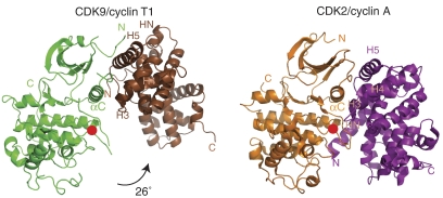
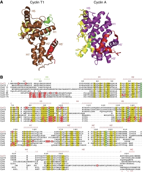
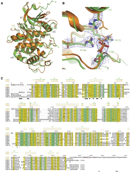
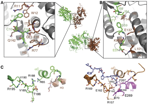
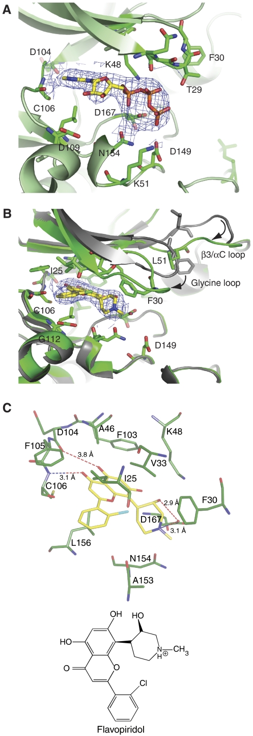
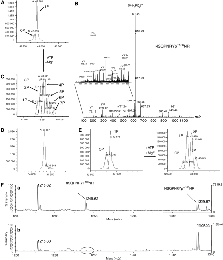
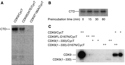
The positive transcription elongation factor b (P-TEFb) (CDK9/cyclin T (CycT)) promotes mRNA transcriptional elongation through phosphorylation of elongation repressors and RNA polymerase II. To understand the regulation of a transcriptional CDK by its cognate cyclin, we have determined the structures of the CDK9/CycT1 and free cyclin T2. There are distinct differences between CDK9/CycT1 and the cell cycle CDK CDK2/CycA manifested by a relative rotation of 26 degrees of CycT1 with respect to the CDK, showing for the first time plasticity in CDK cyclin interactions. The CDK9/CycT1 interface is relatively sparse but retains some core CDK-cyclin interactions. The CycT1 C-terminal helix shows flexibility that may be important for the interaction of this region with HIV TAT and HEXIM. Flavopiridol, an anticancer drug in phase II clinical trials, binds to the ATP site of CDK9 inducing unanticipated structural changes that bury the inhibitor. CDK9 activity and recognition of regulatory proteins are governed by autophosphorylation. We show that CDK9/CycT1 autophosphorylates on Thr186 in the activation segment and three C-terminal phosphorylation sites. Autophosphorylation on all sites occurs in cis.


Figures

Figure 1
Figure 1
The structure of CDK9/CycT1 and comparison with CDK2/CycA. Schematic overall representations of CDK9/CycT1 (left; CDK9 is in green, CycT1 in brown) and CDK2/CycA (right; CDK2 is in orange, CycA in magenta). The phospho-threonine residues in the activation segments are marked by a red circle. The CDK structures have been aligned and the 26° rotation of CycT1 with respect to the position of CycA is indicated. This and other structure figures were prepared with PyMOL (WL DeLano, The PyMOL Molecular Graphics System; DeLano Scientific, San Carlos, CA, USA, 2002).
Figure 2
Figure 2
Comparison of CycT1 and CycA. (A) Schematic diagrams of CycT1 (left) and CycA (right) with secondary structural elements labelled. The colours are the same as Figure 1 but with the HN and HC helices in green and red, respectively, and residues in contact with the CDK in yellow. The orientation of CycA is as in Figure 1. CycT1 has been aligned with CycA for comparison. (B) Structure-based sequence alignment of transcriptional and cell cycle cyclins. Residues conserved in all eight cyclins are in green and those that are similar in yellow. Cyclin A, B, E and T1 residues in contact with their cognate CDK (distance <3.5 Å) are shown highlighted in red. Secondary structure and numbering for CycT1 are shown above the sequences.
Figure 3
Figure 3
CDK9 structure. (A) The structures of CDK9 (green) and CDK2 (orange) superimposed. The cyclins have been omitted for clarity. (B) A close-up of pThr186 in CDK9 with the final 2FoFc electron density and side chain contacts. CDK2 pThr160 and contacts (orange) are superimposed. (C) Sequence comparison of CDK9, CDK2 and other CDKs. Secondary structural elements are indicated for CDK9 and CDK2 in green and orange, respectively. Residues conserved in all CDKs are shown in green and those that are similar in yellow. Residues invariant in CDK9s from human, mouse, chicken, worm, fly and fish but not conserved in the other CDKs are boxed with a red frame in the CDK9 sequence (Supplementary Figure S7). Black circles below the sequences indicate CDK9 residues in contact with CycT1 (<3.5 Å). Red open circles indicate basic residues that contribute to the positively charged surface of CDK9 (Supplementary Figure S4). Black asterisks correspond to autophosphorylation sites.
Figure 4
Figure 4
Details of CDK9/CycT1 interactions. (A) The contacts between CycT1 and the N-terminal residues of CDK9. The view, as indicated in the small overall figure, is 120° rotated about the horizontal axis with respect to Figure 1. Secondary structural elements, shown for guidance but not in contact, are in grey. The colour code is as in Figure 1. (B) The major contacts between CDK9 and CycT1 in the vicinity of the CDK9 αC helix. For further details see text. (C) Selected residues showing the substrate-binding site and phospho-threonine environment for CDK9/CycT1 (left) and CDK2/CycA (right). The substrate heptapeptide bound to CDK2/CycA (Brown et al, 1999) is shown in cyan.
Figure 5
Figure 5
AMPPNP and flavopiridol binding. (A) Details of the interaction of AMPPNP with CDK9/CycT. The CDK9 main chain is represented in green together with the side chains of residues within 3.5 Å of AMPPNP. (B) Details of the interaction of flavopiridol with CDK9/CycT1 and conformational changes. CDK9 in the flavopiridol complex is in green and in the AMPPNP complex in grey. (C) Details of the flavopiridol CDK9 interactions and a schematic representation of flavopiridol. The electron density maps are the final 2FoFc maps contoured at 1σ around the ligand (A) AMPPNP and (B) flavopiridol.
Figure 6
Figure 6
Mass spectrometry analysis of CDK9-FL/CycT1-298 phosphorylation state. (A) CDK9, in the purified CDK9/CycT-298 complex, is ∼80% mono-phosphorylated. The expected MW value of the non-phosphorylated protein is 42800.5. The observed masses are: 0P, 42 803; 1P, 42 881. (B) Phosphorylation is homogeneous on Thr186. MS/MS spectrum of the m/z 665.272+ ion corresponding to CDK9-phosphorylated peptide 179–188. A phosphate loss is observed (m/z 616.292+). Peaks marked with an asterisk correspond to dehydroamino-2-butyric acid containing y″- and b-ions and indicate phosphate location on Thr186 (zoom panel). (C) Distribution of phosphorylation states for CDK9 following autophosphorylation reaction. The observed masses are: 1P, 42 876; 2P, 42 960; 3P, 43 038; 4P, 43 115; 5P, 43 201; 6P, 43 273; 7P, 43 433. (D) Autophosphorylation reaction does not affect CycT1-298 phosphorylation state. The expected MW value for CycT1 34 157 corresponds to the observed mass 34 157. (E) Analysis of autophosphorylated CDK9/CycT1-298 by LC/ESI-MS. Left: starting preparation as purified from insect cells. Right: same preparation following limited in vitro autophosphorylation reaction. The expected MW value of the non-phosphorylated protein is 42 800.5. The observed masses are: 0P, 42 797; 1P, 42 878 (left panel) and 0P, 42 801; 1P, 42 880; 2P, 42 960; 3P, 43 043 (right panel). (F) m/z region (1200–1340) of the MALDI mass spectra from tryptic digests of CDK9 showing the peaks corresponding to peptide 179–188 both in parent and in phosphorylated forms. (a) Digest of CDK9 starting preparation as purified from insect cells. (b) Digest of the same preparation following in vitro autophosphorylation reaction.
Figure 7
Figure 7
Catalytic activity of CDK9/CycT1 on RNA Pol II CTD substrate. (A) CDK9-T186A/CycT1-298 showed no activity on CTD in contrast to CDK9-WT/CycT1-298 or the mutant CDK9-S347E/CycT1-298. (B) CDK9/CycT1-288 activity towards the CTD after preincubation with ATP and removal of excess ATP/ADP. (C) Autophosphorylation reactions for active and kinase-dead CDK9-D167N/CycT1-288 complexes. CDK9-D167N/CycT1-288 or CDK9/CycT1-288 variants as substrates (0.6 μg, (++)) were incubated with or without catalytic amounts (50 ng, (+)) of active CDK 9/CycT1-288.

References

    1. Anand K, Schulte A, Fujinaga K, Scheffzek K, Geyer M (2007) Cyclin box structure of the P-TEFb subunit cyclin T1 derived from a fusion complex with EIAV tat. J Mol Biol 307: 826–836 - PMC - PubMed
    1. Bieniasz PD, Grdina TA, Bogerd HP, Cullen BR (1999) Recruitment of cyclin T1/P-TEFb to an HIV type 1 long terminal repeat promoter proximal RNA target is both necessary and sufficient for full activation of transcription. Proc Natl Acad Sci USA 96: 7791–7796 - PMC - PubMed
    1. Brown NR, Lowe ED, Petri E, Skamnaki V, Antrobus R, Johnson LN (2007) Cyclin B and cyclin A confer different substrate recognition properties on CDK2. Cell Cycle 6: 1350–1359 - PubMed
    1. Brown NR, Noble ME, Endicott JA, Garman EF, Wakatsuki S, Mitchell E, Rasmussen B, Hunt T, Johnson LN (1995) The crystal structure of cyclin A. Structure 3: 1235–1247 - PubMed
    1. Brown NR, Noble ME, Endicott JA, Johnson LN (1999) The structural basis for specificity of substrate and recruitment peptides for cyclin-dependent kinases. Nat Cell Biol 1: 438–443 - PubMed

Publication types