Review
. 2008 Jul;9(7):636-41.
doi: 10.1038/embor.2008.114. Epub 2008 Jun 20.

Polyglutamylation: a fine-regulator of protein function? 'Protein Modifications: beyond the usual suspects' review series

Affiliations
Review

Polyglutamylation: a fine-regulator of protein function? 'Protein Modifications: beyond the usual suspects' review series

Carsten Janke et al. EMBO Rep. 2008 Jul.

Abstract

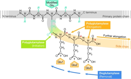
Polyglutamylation is a post-translational modification in which glutamate side chains of variable lengths are formed on the modified protein. It is evolutionarily conserved from protists to mammals and its most prominent substrate is tubulin, the microtubule (MT) building block. Various polyglutamylation states of MTs can be distinguished within a single cell and they are also characteristic of specific cell types or organelles. Polyglutamylation has been proposed to be involved in the functional adaptation of MTs, as it occurs within the carboxy-terminal tubulin tails that participate directly in the binding of many structural and motor MT-associated proteins. The discovery of a new family of enzymes that catalyse this modification has brought new insight into the mechanism of polyglutamylation and now allows for direct functional studies of the role of tubulin polyglutamylation. Moreover, the recent identification of new substrates of polyglutamylation indicates that this post-translational modification could be a potential regulator of diverse cellular processes.


Figures

Figure 1
Figure 1
Mechanism of polyglutamylation. Polyglutamylation takes place on a glutamate residue within the primary sequence of the modified protein (modified Glu). The first reaction, which is initiation (green), generates an amide bond between the γ-carboxyl group of the modified glutamate and the amino group of the first glutamyl unit (Glu1). Subsequent elongation reactions (orange) link the α-carboxyl group of the preceding glutamyl unit with the amino group of the next unit (Glu2, Glu3 and so on). Deglutamylase activity can shorten the side chain. C terminus, carboxy terminus; N terminus, amino terminus.
Figure 2
Figure 2
The occurrence of tubulin polyglutamylation in mammalian cells. (A) Schematic representation of microtubule (MT) species that are modified by polyglutamylation. In proliferating cells, cytoplasmic MTs are polyglutamylated at low levels in interphase. During cell division, polyglutamylation is locally increased on the mitotic spindle and the midbody. Polyglutamylation is also enriched in neurons, as well as on specialized MT structures such as centrioles and axonemes. (B) Parameters of polyglutamylation patterns observed on different MT species in mammals. Values can differ in more distant species. Asterisks indicate that conflicting results about the final length of the side chains have been obtained, probably owing to the methods applied.
Figure 3
Figure 3
Localization of polyglutamylation sites on microtubules. Schematic representation of the arrangement of α–β-tubulin dimer within the microtubule (MT) lattice. The positions of the acidic carboxy-terminal tails of both α- and β-tubulin, which are not resolved in the crystal structure, are indicated in red. Located on the surface of MTs, the tails are the main interaction sites for MT-associated proteins (MAPs) and motors. Polyglutamylation, which occurs on the C-terminal tails, might regulate these interactions. The central panel has been modified with permission from Amos, 2000.
Figure 4
Figure 4
Substrate and reaction preferences of polyglutamylases. (A) Substrate and reaction preferences of known polyglutamylases. Table adapted from van Dijk et al, 2007. *For details of other substrates, see van Dijk et al, 2008. #Enzymatic characteristics deduced from measurements with enriched brain polyglutamylase activity (Janke et al, 2005). (B) Preferences of polyglutamylases determine the distribution of the modification between α- and β-tubulin, and the length of the side chain. Mm, Mus musculus; Tt, Tetrahymena thermophila; TTLL, tubulin tyrosine ligase-like.
None
From left: Krzysztof Rogowski, Juliette van Dijk & Carsten Janke

References

    1. Alexander JE, Hunt DF, Lee MK, Shabanowitz J, Michel H, Berlin SC, MacDonald TL, Sundberg RJ, Rebhun LI, Frankfurter A (1991) Characterization of posttranslational modifications in neuron-specific class III β-tubulin by mass spectrometry. Proc Natl Acad Sci USA 88: 4685–4689 - PMC - PubMed
    1. Amos LA (2000) Focusing-in on microtubules. Curr Opin Struct Biol 10: 236–241 - PubMed
    1. Audebert S, Desbruyeres E, Gruszczynski C, Koulakoff A, Gros F, Denoulet P, Edde B (1993) Reversible polyglutamylation of α- and β-tubulin and microtubule dynamics in mouse brain neurons. Mol Biol Cell 4: 615–626 - PMC - PubMed
    1. Audebert S, Koulakoff A, Berwald-Netter Y, Gros F, Denoulet P, Edde B (1994) Developmental regulation of polyglutamylated α- and β-tubulin in mouse brain neurons. J Cell Sci 107: 2313–2322 - PubMed
    1. Bobinnec Y, Moudjou M, Fouquet JP, Desbruyeres E, Edde B, Bornens M (1998a) Glutamylation of centriole and cytoplasmic tubulin in proliferating non-neuronal cells. Cell Motil Cytoskeleton 39: 223–232 - PubMed

Publication types