Review
. 2008;10(4):291-315.
doi: 10.1007/s12017-008-8044-z. Epub 2008 Jun 20.

Mitochondrial medicine for aging and neurodegenerative diseases

Affiliations
Review

Mitochondrial medicine for aging and neurodegenerative diseases

P Hemachandra Reddy. Neuromolecular Med. 2008.

Abstract

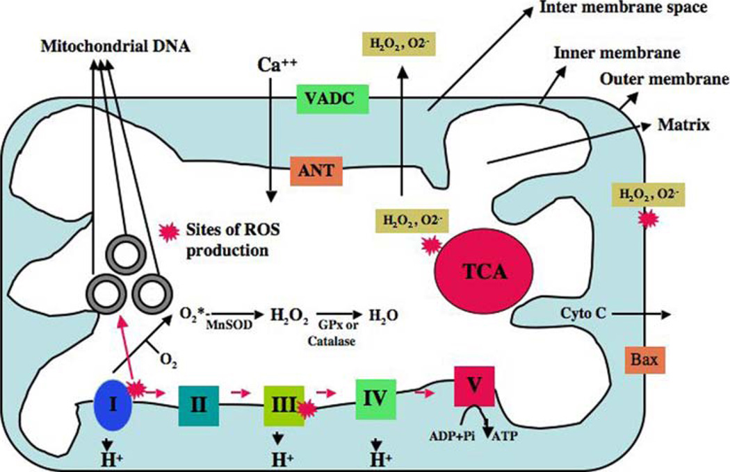
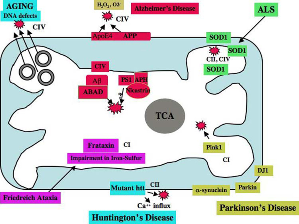
Mitochondria are key cytoplasmic organelles, responsible for generating cellular energy, regulating intracellular calcium levels, altering the reduction-oxidation potential of cells, and regulating cell death. Increasing evidence suggests that mitochondria play a central role in aging and in neurodegenerative diseases, such as Alzheimer's disease, Parkinson's disease, Huntington's disease, amyotrophic lateral sclerosis, and Freidriech ataxia. Further, several lines of evidence suggest that mitochondrial dysfunction is an early event in most late-onset neurodegenerative diseases. Biochemical and animal model studies of inherited neurodegenerative diseases have revealed that mutant proteins of these diseases are associated with mitochondria. Mutant proteins are reported to block the transport of nuclear-encoded mitochondrial proteins to mitochondria, interact with mitochondrial proteins and disrupt the electron transport chain, induce free radicals, cause mitochondrial dysfunction, and, ultimately, damage neurons. This article discusses critical issues of mitochondria causing dysfunction in aging and neurodegenerative diseases, and discusses the potential of developing mitochondrial medicine, particularly mitochondrially targeted antioxidants, to treat aging and neurodegenerative diseases.


Figures

Fig. 1
Fig. 1
Structure of mitochondria and sites of free radical generation. A mitochondrion is compartmentalized with two lipid membranes: the inner mitochondrial membrane and the outer mitochondrial membrane. The inner mitochondrial membrane houses the mitochondrial respiratory chain and provides a highly efficient barrier to ionic flow. Each mitochondrion contains 2–10 copies of a mitochondrial genome. In the ETC complexes I and III leak electrons to oxygen, producing primarily superoxide radicals. Superoxide radicals are dismutated by manganese superoxide dismuase and produce H2O2. In addition, ETC involves H2O2 reducing to H2O and O2 by catalase or glutathione peroxidase accepting electrons donated by NADH and FADH2 and then yielding energy to generate ATP from adenosine diphosphate and inorganic phosphate. Free radicals are also generated by tricarboxylic acid in the matrix. These radicals are carried to the cytoplasm via voltage-dependent anion channels, and may involve oxidation DNA and proteins in the cytoplasm
Fig. 2
Fig. 2
The structure of human mitochondrial DNA. As shown, mitochondrial DNA is circular, and it has 2 strands––a guanine-rich outer strand and cytosine-rich inner strand. The mitochondrial DNA is encoded by 13 polypeptide chains, which encode all essential components of ETC. The mitochondrial DNA also encodes the 22 tRNAs (which are indicated as capital letters outside of outer mitochondrial strand), 12S and 16S rRNA. D-loop represents the control region of mitochondrial DNA. The somatic mtDNA mutations have also been reported to be elevated in Parkinson’s disease (Ikebe et al. 1990; Ozawa et al. 1990), Huntington’s disease (Horton et al. 1995), AD (Corral-Debrinski et al. 1994), livers of alcoholics (Fromenty et al. 1995), ovaries of post-menopausal women (Fromenty et al. 1995), and reduced mobility sperm (Kao et al. 1995)
Fig. 3
Fig. 3
Production of reactive oxygen species and mitochondrial fission. Several mitochondrial toxins, including Aβ peptide, nitric oxide, and rotenone, induce the generation of mitochondrial reactive oxygen species (ROS). In addition, conditions such as hyperglycemia and aging may induce free radicals. The increased production of ROS activates fission molecules, including dynamin-related protein 1 (Drp1) and fission 1 (Fis1), which may lead to mitochondrial fission. Fis 1 protein is localized in the outer mitochondrial membrane, and Drp1 is localized mostly in the cytoplasm, and a fraction of Drp 1 is localized in outer mitochondrial membrane. Drp 1 punctates spots on mitochondria, and these punctate constriction spots lead to mitochondrial fission. The level of mitochondrial fission depends on the free radical production and the activity of Drp1 and Fis1
Fig. 4
Fig. 4
Interaction of proteins in neurodegenerative diseases and mitochondria. The accumulation of mitochondrial DNA mutations may induce ROS production and cause oxidative damage in aged tissues. In AD, age-related production of ROS and decreased ATP levels may contribute to the production of Aβ peptides. Aβ peptides enter mitochondria, induce free radicals, decrease cytochrome oxidase activity, and inhibit ATP generation. In AD brains, APP is transported to outer mitochondrial membranes, blocks the import of nuclear cytochrome oxidase proteins to mitochondria, and may be responsible for decreased cytochrome oxidase activity. In AD neurons (from AD patients, AD transgenic mice, APP cells), Aβ is found in the mitochondrial matrix and binds to ABAD, produces free radicals, and causes mitochondrial dysfunction. The N-terminal portion of ApoE4 is associated with mitochondria, induces free radicals, and causes oxidative damage. Gamma secretase complex proteins, such as presenilins, APH, and nicastrin, were found in the mitochondria and may contribute to Aβ production and free radical generation. In HD neurons, mutant Htt binds to the outer mitochondrial membrane and induces free radical production. H2O2may also interrupt with calcium uptake. In PD neurons, mutant proteins of α-synuclein, parkin, PINK1, and DJ1 are associated with mitochondria and cause mitochondrial dysfunction. Complex I activity is inhibited in PD neurons. In ALS, mutant SOD1 is localized in the inner and outer mitochondrial membranes and matrix, and induces free radical production and oxidative damage. Impairment of complexes II and IV are associated with ALS. Frataxin (a gene product in Freidriech ataxia) is a mitochondrial protein responsible for heme biosynthesis and the formation of iron-sulfur clusters. In Freidriech ataxia, mutant frataxin facilitates the accumulation of iron in mitochondria and induces free radicals
Fig. 5
Fig. 5
Strategies of neuronal protection in aging and neurodegenerative diseases. 1. A generic mitochondrially targeted antioxidant is shown constructed by the covalent attachment of an antioxidant molecule to the lipophilic triphenylphosphonium cation. Antioxidant molecules accumulate 5–10 fold in the cytoplasm, which is driven by plasma membrane potential, and then further accumulates 100–500 fold in the mitochondria. Mitochondrial antioxidants rapidly neutralize free radicals and reduce mitochondrial toxicity. 2. Glutathione is synthesized in the cytoplasm and transported to mitochondria. N-acetyl-L-cysteine provides s cysteine for glutathione synthase. Choline esters of glutathione and N-acetyl-L-cysteine have been used to increase mitochondrial glutathione and N-acetyl-L-cysteine. Increased glutathione can rapidly neutralize free radicals and protect mitochondria from oxidative insults. 3. SS peptides are cell-permeable, mitochondrially targeted antioxidants that are reported to protect mitochondria from oxidative damage. These SS peptides have a sequence motif that allows them to target mitochondria several hundred fold more than natural antioxidants. Once SS peptides reach mitochondria, the SS peptides rapidly neutralize free radicals and decrease mitochondrial toxicity
Fig. 6
Fig. 6
Neuroprotective effects of MitoQ and SS-31. N2a cells were grown in a serum-free medium for 7 days and treated with MitoQ (0.3 µM) and SS-31 (0.1nM). After 48 h of treatment, neurite outgrowth was examined after immunostaining with an anti-dynamin-related protein 1 or Drp1 (enriched in neurons, particularly at synapses). As shown, increased neurite outgrowth has been observed in neurons treated with MitoQ and SS-31, in contrast to untreated N2a cells. This observation indicates that mitochondrially targeted antioxidants at very low concentrations are cytoprotective, and may have a role in neurite outgrowth and synaptic connectivity
Fig. 7
Fig. 7
Neuronal protection of calorie-restricted diet and Sirtuins in aging and neurodegenerative diseases

References

    1. Abe Y, Hashimoto Y, Tomita Y, Terashita K, Aiso S, Tajima H, et al. Cytotoxic mechanisms by M239 V presenilin 2, a little-analyzed Alzheimer’s disease-causative mutant. Journal of Neuroscience Research. 2004;77:583–595. - PubMed
    1. Abeliovich A, Beal MF. Parkinsonism genes: Culprits and clues. Journal of Neurochemistry. 2006;99:1062–1072. - PubMed
    1. Afifi AK, Aleu FP, Goodgold J, MacKay B. Ultrastructure of atrophic muscle in amyotrophic lateral sclerosis. Neurology. 1966;16:475–481. - PubMed
    1. Anandatheerthavarada HK, Biswas G, Robin MA, Avadhani NG. Mitochondrial targeting and a novel transmembrane arrest of Alzheimer’s amyloid precursor protein impairs mitochondrial function in neuronal cells. Journal of Cell Biology. 2003;161:41–54. - PMC - PubMed
    1. Andersen JK. Iron dysregulation and Parkinson’s disease. Journal of Alzheimer’s Disease. 2004;6:S47–S52. - PubMed

Publication types

MeSH terms