

Muscle and bone plasticity after spinal cord injury: review of adaptations to disuse and to electrical muscle stimulation
- PMID: 18566946
- PMCID: PMC2744487
- DOI: 10.1682/jrrd.2007.02.0031
Muscle and bone plasticity after spinal cord injury: review of adaptations to disuse and to electrical muscle stimulation
Abstract
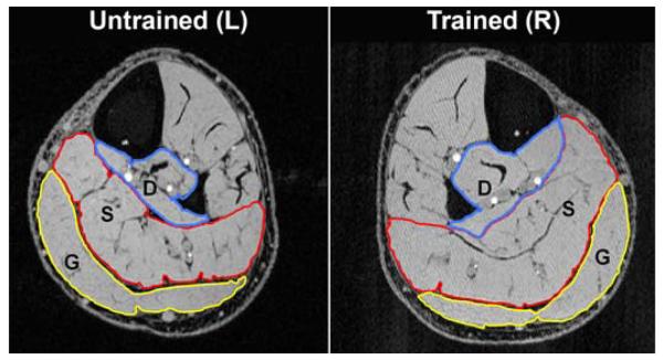
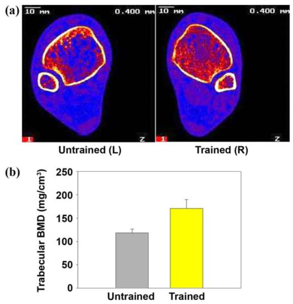
The paralyzed musculoskeletal system retains a remarkable degree of plasticity after spinal cord injury (SCI). In response to reduced activity, muscle atrophies and shifts toward a fast-fatigable phenotype arising from numerous changes in histochemistry and metabolic enzymes. The loss of routine gravitational and muscular loads removes a critical stimulus for maintenance of bone mineral density (BMD), precipitating neurogenic osteoporosis in paralyzed limbs. The primary adaptations of bone to reduced use are demineralization of epiphyses and thinning of the diaphyseal cortical wall. Electrical stimulation of paralyzed muscle markedly reduces deleterious post-SCI adaptations. Recent studies demonstrate that physiological levels of electrically induced muscular loading hold promise for preventing post-SCI BMD decline. Rehabilitation specialists will be challenged to develop strategies to prevent or reverse musculoskeletal deterioration in anticipation of a future cure for SCI. Quantifying the precise dose of stress needed to efficiently induce a therapeutic effect on bone will be paramount to the advancement of rehabilitation strategies.
Figures
References
-
- Stover SL, DeLisa JA, Whiteneck GG. Spinal cord injury: Clinical outcomes from the model systems. Aspen Publishers, Inc; Gaithersburg (MD): 1995.
-
- Wilmet E, Ismail AA, Heilporn A, Welraeds D, Bergmann P. Longitudinal study of the bone mineral content and of soft tissue composition after spinal cord section. Paraplegia. 1995;33(11):674–77. [PMID: 8584304] - PubMed
-
- Abramson AS. Bone disturbances in injuries to the spinal cord and cauda equina (paraplegia) J Bone Joint Surg. 1948;30:982–87. - PubMed
-
- Claus-Walker J, Carter RE, Campos RJ, Spencer WA. Sitting, muscular exercises, and collagen metabolism in tetraplegia. Am J Phys Med. 1979;58(6):285–93. [PMID: 517641] - PubMed
Publication types
MeSH terms
Grants and funding
LinkOut - more resources
Full Text Sources
Medical